May-Thurner syndrome and iliac vein stenting: video explainer
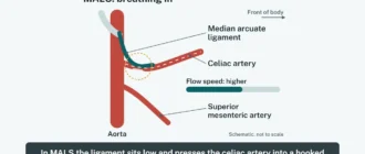
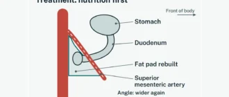
A short captioned video, about a minute and a half, with no sound. It shows how the right iliac artery can squeeze the left iliac vein, why that raises the risk of a clot in the left leg, and how a stent is placed, step by step.
Chapters
- Normal anatomy
- The squeeze
- Slow flow, clot risk
- A stent holds it open
- Stenting: Access through a vein
- Stenting: Imaging the narrowing
- Stenting: Balloon and stent
- Stenting: After the procedure
Full transcript
- 0:03 The two iliac veins join to form the main vein of the belly, the inferior vena cava.
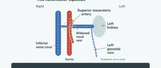
- 0:10 The left iliac vein passes behind the right iliac artery, in front of the spine.
- 0:16 In May-Thurner syndrome, the artery presses the vein against the spine.
- 0:20 A squeeze alone is common: one CT study found it in 24% of people without vein symptoms.
- 0:27 Blood from the left leg drains less easily and can pool.
- 0:32 This raises the risk of a clot in a deep leg vein (DVT).
- 0:37 Left leg swelling and pain are common symptoms.
- 0:40 A stent, a mesh tube inside the vein, holds it open.
- 0:44 In one study, 96% to 99% of stents stayed open over a median of 42 months.
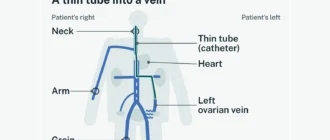
- 0:54 Stenting, step 1: a vein in the leg is punctured with ultrasound guidance.
- 0:59 A thin tube (sheath) lets the tools pass up to the narrowing.
- 1:04 Step 2: dye X-rays (venography) and a tiny ultrasound probe inside the vein (IVUS) measure the narrowing.
- 1:11 In one large study, IVUS found over 90% of these narrowings, venography 66%.
- 1:16 Step 3: a balloon widens the narrow part.
- 1:19 A self-expanding stent is left in place to hold the vein open.
- 1:24 Afterwards, doctors may prescribe blood thinners; there is no agreed best plan yet.
- 1:29 Ultrasound checks follow to see that the stent stays open.
About this video
- The drawings are schematic and not to scale. Each step is based on published research; the sources are listed with the same steps in our May-Thurner syndrome guide and on the stent results page.
- Numbers in the video come from single studies and are examples, not predictions for one person.
- This video is educational only and is not medical advice. Talk with your doctor about your own situation.
See also: how often the stent stays open in every published study and US hospitals that publish on May-Thurner syndrome.
MALS Compass is an independent educational website, not a medical provider, and is not affiliated with any hospital, clinic or university. This page is educational information and does not replace advice from your own doctor. For any question about your health, please contact your doctor or an official healthcare institution.