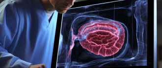

Diagnosing MALS: The Role of Doppler Ultrasound, CT Angiography, and Other Imaging Studies.
MALS (Median Arcuate Ligament Syndrome) is a rare but debilitating vascular disorder that occurs when the median arcuate ligament compresses the celiac artery, leading to chronic abdominal pain and other symptoms. Diagnosing MALS can be challenging, as its symptoms are often nonspecific and overlap with those of other gastrointestinal disorders. Therefore, accurate and timely diagnosis is crucial in order to provide effective treatment and improve the quality of life for patients.
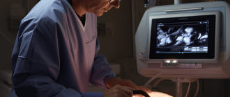
Doppler ultrasound and CT angiography play a critical role in the diagnosis of MALS. Doppler ultrasound is a non-invasive imaging technique that uses sound waves to measure the blood flow in the abdominal arteries. It can detect any abnormalities in the celiac artery, such as stenosis or turbulence caused by the compression of the median arcuate ligament. Doppler ultrasound is especially useful in initial screening and can help determine whether further imaging tests are needed.
CT angiography is another imaging technique that uses a combination of x-rays and contrast dye to create detailed images of the blood vessels. It provides a more comprehensive view of the celiac artery and its surrounding structures, allowing for a more accurate assessment of the extent of compression and its effects on blood flow. CT angiography can also help identify any other potential causes of abdominal pain, such as aneurysms or tumors.
In addition to Doppler ultrasound and CT angiography, other imaging techniques, such as magnetic resonance angiography (MRA) and digital subtraction angiography (DSA), may also be used in the diagnosis of MALS. However, these techniques are usually reserved for cases where the initial imaging tests are inconclusive, or when there is a need for more detailed visualization of the celiac artery and its branches.
In conclusion, the role of doppler ultrasound, CT angiography, and other imaging techniques in the diagnosis of MALS cannot be overstated. These non-invasive and minimally invasive procedures provide valuable information about the condition of the celiac artery and its blood flow, helping clinicians make an accurate diagnosis and develop a targeted treatment plan for patients with MALS.
Understanding MALS: What Is It and How Does It Affect the Body?
MALS, or Median Arcuate Ligament Syndrome, is a rare condition that affects the blood vessels in the abdomen. It occurs when the median arcuate ligament, a band of tissue that connects the diaphragm to the spine, compresses the celiac artery and surrounding blood vessels. This compression can restrict blood flow to the abdominal organs and cause a variety of symptoms.
The role of imaging techniques like Doppler ultrasound and CT angiography is crucial in diagnosing MALS. Doppler ultrasound uses sound waves to create images of blood flow, allowing doctors to visualize any abnormalities or blockages in the arteries. CT angiography, on the other hand, involves injecting a contrast dye into the bloodstream and taking detailed X-ray images to identify any abnormalities in the blood vessels.
Patients with MALS may experience a wide range of symptoms, including severe abdominal pain after eating, weight loss, nausea, and vomiting. The compression of the celiac artery can lead to decreased blood flow to the stomach, liver, and other organs, causing these symptoms. In some cases, MALS may also cause abnormal bruising or discoloration of the abdomen.
Treatment for MALS typically involves surgery to release the compression on the celiac artery and improve blood flow. This procedure, known as a laparoscopic decompression, involves cutting the median arcuate ligament to relieve the pressure on the blood vessels. In some cases, additional surgical interventions may be needed to address any complications or underlying conditions.
It is important for individuals experiencing symptoms of MALS to seek medical attention and undergo proper diagnostic testing. Imaging techniques like Doppler ultrasound and CT angiography play a vital role in accurately diagnosing this condition and determining the most appropriate treatment plan. Early diagnosis and intervention can help improve quality of life for individuals affected by MALS.
Key Symptoms of MALS: Identifying the Red Flags
The role of diagnosing MALS involves recognizing the key symptoms that may indicate the presence of this condition. Medical professionals rely on various imaging techniques, such as Doppler ultrasound and CT angiography, to aid in the diagnosis.
Some key symptoms that may raise red flags for MALS include:
- Chronic abdominal pain: Patients with MALS often experience chronic, severe abdominal pain that may be worsened by eating. The pain is typically located in the upper abdomen and may be described as constant or intermittent.
- Weight loss: Unexplained weight loss can be a symptom of MALS. This is often due to a reduced appetite and difficulty eating, as the compression of the celiac artery can interfere with proper digestion and nutrient absorption.
- Nausea and vomiting: Some individuals with MALS may experience recurring episodes of nausea and vomiting, especially after meals.
- Postprandial fullness: Many MALS patients report a feeling of fullness or discomfort after eating, even when consuming small amounts of food.
- Diarrhea: Frequent bowel movements or diarrhea can be a symptom of MALS, as the compromised blood flow to the intestines can affect their normal function.
In addition to these key symptoms, patients may also present with other signs, such as fatigue, malnutrition, and anemia. It is important for medical professionals to carefully evaluate these symptoms and consider the possibility of MALS when making a diagnosis.
The Role of Doppler Ultrasound in Diagnosing MALS
Doppler ultrasound plays a crucial role in the diagnosis of Median Arcuate Ligament Syndrome (MALS). MALS is a condition where the median arcuate ligament, located near the diaphragm, compresses the celiac artery and causes symptoms such as abdominal pain, weight loss, and digestive issues. To confirm a diagnosis of MALS, imaging techniques like Doppler ultrasound, CT angiography, and magnetic resonance angiography (MRA) are often used.
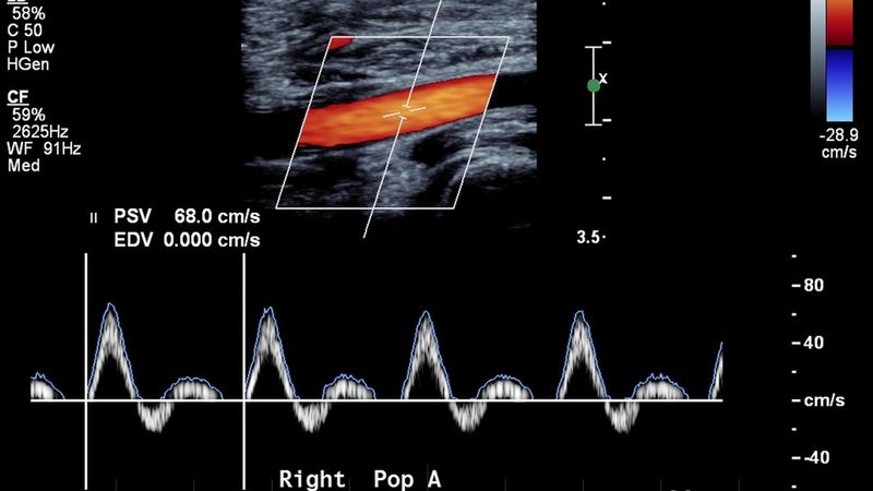
Doppler ultrasound is a non-invasive imaging technique that uses sound waves to create real-time images of blood flow in the body. This technique is particularly useful in diagnosing MALS because it allows healthcare providers to assess the blood flow in the celiac artery and identify any abnormalities or blockages caused by the compression of the median arcuate ligament.
During a Doppler ultrasound examination, a transducer is placed on the abdominal area, specifically targeting the celiac artery. The transducer emits sound waves that bounce off the moving blood cells in the artery. By analyzing the frequency changes of these sound waves, healthcare providers can determine the speed and direction of blood flow in the celiac artery.
In patients with MALS, Doppler ultrasound can reveal reduced blood flow or turbulence in the celiac artery. These findings, combined with a patient’s symptoms and medical history, can help healthcare providers make an accurate diagnosis of MALS.
It is important to note that while Doppler ultrasound is a valuable tool in diagnosing MALS, it is often used in conjunction with other imaging techniques, such as CT angiography or MRA. These additional imaging techniques provide more detailed anatomical information and can confirm the presence of other potential causes of symptoms, such as vascular abnormalities or tumors.
In conclusion, Doppler ultrasound plays a crucial role in the diagnosis of MALS by allowing healthcare providers to assess blood flow in the celiac artery. This non-invasive imaging technique helps identify any abnormalities or blockages caused by the compression of the median arcuate ligament. However, it is important to use Doppler ultrasound in conjunction with other imaging techniques for a comprehensive evaluation and accurate diagnosis of MALS.
CT Angiography: A Comprehensive Imaging Technique for MALS Diagnosis
The role of imaging techniques, such as Doppler ultrasound, CT angiography, and others, is crucial in diagnosing median arcuate ligament syndrome (MALS), a condition characterized by compression of the celiac artery by the median arcuate ligament.
Among these imaging techniques, CT angiography plays a significant role in providing a comprehensive evaluation of MALS.
CT angiography combines the use of a contrast agent and computed tomography to visualize the celiac artery and surrounding structures. It provides detailed images that aid in the accurate identification and measurement of the median arcuate ligament, as well as the degree of stenosis or compression of the celiac artery.
During the procedure, the patient is positioned on a table that slides into the CT scanner. The contrast agent is injected into the patient’s veins to enhance the visibility of blood vessels. The scanner then takes multiple X-ray images, which are reconstructed to create a detailed 3D representation of the celiac artery and surrounding anatomy.
This noninvasive imaging technique allows for visualization of the celiac artery and its branches, as well as the nearby organs and tissues. It can identify any abnormalities, such as narrowing or blockages in the celiac artery, which may indicate the presence of MALS.
In addition to providing detailed anatomical information, CT angiography can also assess blood flow dynamics in the celiac artery. The use of contrast agents enables the evaluation of blood flow patterns and velocities, which can help in identifying abnormalities in blood flow caused by compression of the celiac artery.
Overall, CT angiography is a valuable tool in the diagnosis of MALS. Its ability to provide detailed anatomical and functional information makes it an essential imaging technique for evaluating patients suspected of having MALS. Its noninvasiveness, accuracy, and comprehensiveness make it an optimal choice for diagnosing this condition.
MRI: An Alternative Imaging Option for MALS Evaluation
While Doppler ultrasound and CT angiography are commonly used imaging techniques for diagnosing median arcuate ligament syndrome (MALS), MRI can also play a role in evaluating this condition.
MALS, a vascular disorder characterized by compression of the celiac artery by the median arcuate ligament of the diaphragm, can cause chronic abdominal pain and other symptoms. The diagnosis of MALS is often challenging due to its nonspecific symptoms and the need for imaging studies to confirm the diagnosis.
Doppler ultrasound and CT angiography are frequently used to assess blood flow and identify anatomical abnormalities in patients suspected to have MALS. Doppler ultrasound uses sound waves to measure the velocity and direction of blood flow, while CT angiography combines computed tomography and angiography to obtain detailed images of blood vessels.
However, MRI can offer additional advantages in the evaluation of MALS. MRI provides excellent soft tissue contrast and can visualize the celiac artery and surrounding structures in multiple planes. It can also assess blood flow dynamics and detect collateral vessels, which may be present in patients with MALS.
An MRI examination for MALS typically involves a combination of different sequences, such as T1-weighted, T2-weighted, and contrast-enhanced images. These sequences allow for a comprehensive assessment of the celiac artery, surrounding structures, and any potential abnormalities or narrowing of the vessel.
In addition to its diagnostic capabilities, MRI is non-invasive and does not involve exposure to ionizing radiation, unlike CT angiography. This makes MRI a safer imaging option, especially for younger patients or those who may require repeated imaging studies.
In conclusion, while Doppler ultrasound and CT angiography are commonly used for diagnosing MALS, MRI can serve as an alternative imaging option. MRI provides detailed anatomical and functional information, without the use of ionizing radiation. Its use in the evaluation of MALS can help clinicians make an accurate diagnosis and develop an appropriate treatment plan for patients with this challenging condition.
Exploring the Use of Digital Subtraction Angiography in MALS Diagnosis
Introduction:
Diagnosing Median Arcuate Ligament Syndrome (MALS) can be challenging due to its nonspecific symptoms. Various imaging techniques, such as ultrasound, CT angiography, and Doppler ultrasound, are commonly used to aid in the diagnosis. In recent years, digital subtraction angiography (DSA) has emerged as an advanced imaging technique that provides detailed visualization of the arterial and venous anatomy. This article explores the use of DSA in diagnosing MALS.
What is Digital Subtraction Angiography (DSA)?
- DSA is a radiographic technique that allows for the visualization of blood vessels using contrast media.
- It involves taking a series of X-ray images before and after injecting a contrast agent into the bloodstream.
- The pre-injection images are then subtracted from the post-injection images, resulting in a clear visualization of the blood vessels.
- DSA provides high-resolution images that can reveal detailed information about vascular anatomy, including any compression or stenosis of the arteries.
Advantages of DSA in MALS Diagnosis:
- Accurate Visualization: DSA allows for accurate visualization of the celiac artery and its branches, which is crucial in diagnosing MALS. It provides detailed information about any compression or obstruction of the vessels.
- Dynamic Imaging: DSA can capture real-time images of blood flow, providing dynamic information about the arterial and venous circulation. This can help in identifying abnormal blood flow patterns associated with MALS.
- Guidance for Interventional Procedures: DSA can also be used as a guidance tool during interventional procedures, such as angioplasty or stenting, to treat MALS. The detailed visualization provided by DSA helps in accurate placement of the devices.
Limitations of DSA:
- DSA is an invasive procedure that requires the insertion of a catheter into the blood vessels.
- There is a risk of complications, such as bleeding or infection, associated with the insertion of the catheter.
- Additionally, DSA exposes the patient to ionizing radiation, which may have harmful effects.
Conclusion:
While ultrasound, CT angiography, and Doppler ultrasound continue to be the primary imaging techniques in diagnosing MALS, DSA offers a valuable adjunct in certain cases. Its ability to provide accurate visualization of the arterial and venous anatomy, dynamic imaging capabilities, and guidance during interventional procedures make DSA a promising tool in MALS diagnosis and treatment. However, the invasive nature and potential risks associated with DSA should be carefully considered before its use.
Non-Invasive Imaging Techniques for MALS Diagnosis
Malnutritive Lymphatic and Syndromic (MALS) disease is a condition that affects the blood supply to the mesenteric arteries, leading to chronic abdominal pain and other gastrointestinal symptoms. In diagnosing MALS, non-invasive imaging techniques play a crucial role.
Doppler ultrasound is one such imaging technique that can be used to diagnose MALS. It utilizes sound waves to create images of blood flow through the arteries. By analyzing the blood flow in the mesenteric arteries, doctors can identify any abnormalities or restrictions in the blood supply, which can indicate the presence of MALS.
CT angiography is another non-invasive imaging technique that can aid in diagnosing MALS. It involves injecting a contrast dye into the patient’s bloodstream and then taking a series of X-ray images. The dye helps highlight the blood vessels, allowing doctors to assess the blood flow and detect any abnormalities.
Other imaging techniques, such as magnetic resonance angiography (MRA) and computed tomography (CT) scans, can also be used in the diagnosis of MALS. These techniques provide detailed images of the blood vessels and allow doctors to assess the blood flow and identify any obstructions or abnormalities.
The role of non-invasive imaging techniques in diagnosing MALS is crucial. These techniques help doctors visualize the blood vessels and assess the blood flow, allowing for an accurate diagnosis of the condition. By utilizing Doppler ultrasound, CT angiography, MRA, and CT scans, physicians can identify MALS and develop an appropriate treatment plan for patients suffering from this condition.
The Importance of Combining Different Imaging Modalities in MALS Diagnosis
Diagnosing Median Arcuate Ligament Syndrome (MALS) involves the use of various imaging techniques, including Doppler ultrasound, CT angiography, and other modalities. Each of these imaging techniques plays a crucial role in accurately diagnosing MALS and determining the appropriate treatment plan.
Doppler Ultrasound:
Doppler ultrasound is often one of the initial imaging techniques used for diagnosing MALS. It is a non-invasive procedure that evaluates blood flow through the vessels in the abdomen. Doppler ultrasound can help identify any abnormalities or blockages in the celiac artery, which is often compressed in patients with MALS.
CT Angiography:
CT angiography is another important imaging modality for diagnosing MALS. This technique involves the injection of a contrast dye into the patient’s bloodstream, followed by a series of detailed X-ray images. CT angiography provides a high-resolution visualization of the blood vessels, allowing for the detection of any narrowing or compression in the celiac artery.
Role of Other Imaging Techniques:
In addition to Doppler ultrasound and CT angiography, other imaging techniques may also be used in the diagnosis of MALS. These may include magnetic resonance imaging (MRI), magnetic resonance angiography (MRA), or even mesenteric digital subtraction angiography (DSA). These additional imaging techniques can provide further insights into the extent of compression or any associated vascular abnormalities.
Benefits of Combining Modalities:
By combining different imaging modalities, healthcare professionals can obtain a comprehensive evaluation of the celiac artery and surrounding structures. This multimodal approach allows for a more accurate diagnosis of MALS and can help guide treatment decisions. For example, Doppler ultrasound can provide real-time information on blood flow, while CT angiography can offer detailed anatomical images.
Conclusion:
Diagnosing MALS requires the use of multiple imaging modalities, including Doppler ultrasound, CT angiography, and possibly others. The combination of these techniques plays a vital role in accurately diagnosing MALS and determining appropriate treatment strategies. The multimodal approach ensures a comprehensive evaluation of the celiac artery and aids in making informed clinical decisions.
Surgical Planning: How Imaging Techniques Aid in Determining the Best Treatment Approach for MALS
When it comes to diagnosing and treating median arcuate ligament syndrome (MALS), imaging techniques play a crucial role in surgical planning. These techniques, such as doppler ultrasound and CT angiography, provide valuable insights into the anatomy and blood flow of the affected vessels, helping determine the best treatment approach for each individual case.
Doppler ultrasound is often the initial imaging modality used to evaluate patients with suspected MALS. It is a non-invasive test that uses sound waves to assess blood flow through the arteries and veins. In MALS, doppler ultrasound can detect abnormalities in the celiac artery, such as compression or stenosis caused by the median arcuate ligament. This information is essential in confirming the diagnosis and determining the severity of the condition.
CT angiography is another imaging technique that provides detailed images of the blood vessels in the abdomen. It involves injecting a contrast dye into the bloodstream and using a CT scanner to capture images of the dye as it moves through the vessels. CT angiography can help visualize the compression of the celiac artery by the median arcuate ligament and assess the extent of the narrowing. It also allows the surgeon to evaluate the collateral blood vessels that may have developed as a compensatory mechanism.
The information obtained from these imaging techniques is crucial in determining the best treatment approach for MALS patients. In some cases, conservative management, such as dietary modifications and lifestyle changes, may be sufficient to alleviate symptoms. However, for patients with more severe compression or persistent symptoms, surgical intervention may be necessary.
Surgical planning for MALS often involves a multidisciplinary team, including vascular surgeons, radiologists, and gastroenterologists. The imaging findings guide the surgical team in selecting the most appropriate procedure to relieve the compression of the celiac artery. The goal of the surgery is to restore proper blood flow to the digestive organs and alleviate symptoms.
In conclusion, imaging techniques, such as doppler ultrasound and CT angiography, play a critical role in the surgical planning for MALS. They provide valuable insights into the anatomy and blood flow of the affected vessels, helping determine the severity of the condition and guide the selection of the most appropriate treatment approach. By using these imaging techniques, healthcare professionals can ensure that MALS patients receive the best possible care tailored to their individual needs.
Imaging-Guided Interventions for MALS: A Minimally Invasive Treatment Option
When diagnosing median arcuate ligament syndrome (MALS), imaging techniques play a crucial role in identifying the condition and determining the appropriate treatment options. While Doppler ultrasound and CT angiography are commonly used for the initial diagnosis of MALS, they also serve a vital purpose in guiding treatment interventions.
Doppler Ultrasound:
Doppler ultrasound uses sound waves to produce images and measure blood flow within the arteries and veins. In the context of MALS, Doppler ultrasound can help identify the characteristic narrowing or compression of the celiac artery caused by the median arcuate ligament, confirming the diagnosis.
CT Angiography:
CT angiography is a diagnostic technique that combines the use of a contrast dye and CT scanning to visualize blood vessels in detail. With MALS, CT angiography can reveal the compression of the celiac artery and provide a clear image of the surrounding anatomy, helping to rule out other potential causes of abdominal pain.
While these imaging techniques are essential for diagnosing MALS, they also play a crucial role in guiding minimally invasive treatment options.
Minimally Invasive Treatment Options:
Once MALS is diagnosed, imaging-guided interventions can be performed to relieve the compression of the celiac artery and alleviate symptoms. These interventions typically involve the use of endovascular techniques such as angioplasty and stenting.
During angioplasty, a thin catheter is inserted into the affected blood vessel, and a balloon at the tip of the catheter is inflated to widen the narrowed artery. This helps restore normal blood flow and alleviate symptoms caused by the compression of the celiac artery.
In some cases, a stent may also be placed during the procedure to help maintain the widened artery. A stent is a small metal mesh tube that is inserted into the treated artery to provide structural support and prevent re-narrowing.
Imaging techniques, such as Doppler ultrasound and CT angiography, are used to guide these interventions by providing real-time visualization of the blood vessels during the procedure. This allows the interventional radiologist to precisely position the catheter, balloon, and stent, ensuring optimal results.
In conclusion, imaging-guided interventions play a crucial role in the management of MALS. By utilizing techniques such as Doppler ultrasound and CT angiography, interventional radiologists are able to accurately diagnose MALS and guide minimally invasive treatment options to provide relief for patients suffering from this condition.
Case Studies: Real-Life Examples Demonstrating the Diagnostic Value of Imaging Techniques in MALS
Diagnosing Median Arcuate Ligament Syndrome (MALS) can be challenging as its symptoms can mimic other abdominal conditions. However, imaging techniques such as CT angiography and Doppler ultrasound play a crucial role in accurately diagnosing MALS. Below are some real-life case studies highlighting the diagnostic value of these imaging techniques in MALS.
-
Case Study 1: A 45-year-old female presented with chronic abdominal pain and weight loss. Initial clinical examination and laboratory tests were inconclusive. However, a Doppler ultrasound showed increased blood flow velocities in the celiac artery during expiration, indicating compression of the vessel by the median arcuate ligament. CT angiography further confirmed the diagnosis, revealing significant narrowing of the celiac artery at the level of the median arcuate ligament. Surgical intervention was performed, and the patient experienced significant relief from her symptoms.
-
Case Study 2: A 32-year-old male complained of postprandial abdominal pain and unintentional weight loss. Initial workup included laboratory tests and endoscopy, which were unremarkable. However, a CT angiography revealed compression of the celiac artery by the median arcuate ligament. Doppler ultrasound was performed to assess the severity of stenosis, and it showed abnormal flow velocities in the celiac artery consistent with MALS. The patient underwent surgical release of the median arcuate ligament and reported complete resolution of his symptoms postoperatively.
-
Case Study 3: A 50-year-old female presented with recurrent abdominal pain and early satiety. Clinical examination and laboratory tests were inconclusive. To further evaluate her symptoms, a Doppler ultrasound and CT angiography were performed. The Doppler ultrasound demonstrated elevated blood flow velocities in the celiac artery during forced expiration, suggesting compression by the median arcuate ligament. CT angiography confirmed stenosis of the celiac artery at the level of the ligament. Laparoscopic release of the ligament was performed, resulting in significant improvement of the patient’s symptoms.
These case studies demonstrate the immense diagnostic value of imaging techniques, such as Doppler ultrasound and CT angiography, in diagnosing MALS. By accurately identifying the compression of the celiac artery by the median arcuate ligament, these imaging techniques play a crucial role in determining the most appropriate treatment strategies for patients suffering from MALS.
Limitations of Imaging Techniques in MALS Diagnosis: What You Need to Know
When it comes to diagnosing Median Arcuate Ligament Syndrome (MALS), a condition characterized by the compression of the celiac artery by the median arcuate ligament, imaging techniques play a crucial role. However, it is important to understand that these imaging techniques have their limitations and may not always provide a definitive diagnosis. Here are some limitations to be aware of:
- Ultrasound: While ultrasound can provide valuable information about blood flow and detect any abnormalities in the celiac artery, it may not always be able to clearly visualize the exact cause of the compression. The interpretation of ultrasound images can also be subjective, depending on the expertise of the operator.
- Doppler Ultrasound: Doppler ultrasound uses sound waves to evaluate blood flow through the celiac artery. While it can help identify abnormal velocities indicative of MALS, it cannot provide a complete anatomical picture of the ligament and its interaction with surrounding structures.
- CT Angiography: CT angiography is often considered the gold standard for diagnosing MALS. It provides detailed images of the celiac artery and surrounding structures, allowing for a more accurate assessment. However, it does involve the use of radiation and contrast dye, which may not be suitable for all patients, especially those with kidney problems or allergies to contrast agents.
It is important for medical professionals to interpret imaging findings in the context of the patient’s clinical presentation and symptoms. Other diagnostic methods, such as clinical evaluation, medical history, and exclusion of other possible causes, may also be necessary to confirm a diagnosis of MALS.
Furthermore, it is worth noting that imaging techniques can only provide a snapshot of the condition at a specific point in time. MALS is a dynamic disease, and the severity of compression may vary with different body positions and respiratory phases. Therefore, multiple imaging studies may be required to accurately assess the extent of the compression and its impact on blood flow.
In conclusion, while imaging techniques like ultrasound and CT angiography play an important role in diagnosing MALS, they have their limitations. It is important for healthcare professionals to be aware of these limitations and use a comprehensive approach to diagnose and manage MALS effectively.
The Future of Imaging Techniques in MALS Diagnosis: Innovations and Advancements
Doppler ultrasound and CT angiography have played a crucial role in diagnosing MALS (Median Arcuate Ligament Syndrome). These imaging techniques have allowed clinicians to visualize the blood flow to the celiac artery and identify any abnormalities or compression in the area. However, the field of diagnostic imaging is constantly evolving, and new innovations and advancements are being made that could further improve the diagnosis of MALS.
One exciting area of development is the use of 3D imaging techniques. While traditional ultrasound and CT angiography provide two-dimensional images, 3D imaging allows for a more comprehensive view of the celiac artery and surrounding structures. This enhanced visualization can help clinicians better understand the anatomy and pathology of MALS and improve diagnostic accuracy.
Another promising advancement is the use of contrast-enhanced ultrasound (CEUS). CEUS involves injecting a contrast agent into the bloodstream, which highlights blood vessels and improves the visualization of blood flow. This technique can provide additional information about the hemodynamics of the celiac artery and may help differentiate between MALS and other conditions with similar symptoms.
Magnetic Resonance Imaging (MRI) is another imaging technique that shows promise in the diagnosis of MALS. MRI can provide detailed images of the celiac artery and surrounding tissues without the use of ionizing radiation. Additionally, functional MRI techniques can assess blood flow and tissue perfusion, providing valuable information about the vascular dynamics in MALS patients.
Furthermore, advancements in image processing and analysis software have the potential to enhance the diagnostic capabilities of existing imaging techniques. These software tools can automate the measurement of blood flow velocities, identify areas of stenosis or compression, and aid in the interpretation of imaging data. This can help clinicians make more accurate and timely diagnoses, improving patient outcomes.
In conclusion, the future of imaging techniques in MALS diagnosis holds great promise. Innovations and advancements such as 3D imaging, contrast-enhanced ultrasound, MRI, and image processing software have the potential to enhance diagnostic accuracy and improve patient care. As these technologies continue to develop, it is likely that the diagnosis of MALS will become even more precise and efficient, leading to better treatment outcomes for patients suffering from this condition.
Q&A:
What is MALS?
MALS stands for Median Arcuate Ligament Syndrome, a condition in which the median arcuate ligament, which is a fibrous band of tissue, compresses the celiac artery and the surrounding nerves.
How is MALS diagnosed?
MALS can be diagnosed using a variety of imaging techniques, including Doppler ultrasound, CT angiography, and magnetic resonance angiography (MRA). These techniques allow doctors to visualize the anatomy of the celiac artery and determine if there is any compression or narrowing.
What is the role of Doppler ultrasound in diagnosing MALS?
Doppler ultrasound is a non-invasive imaging technique that uses sound waves to measure blood flow. In the case of MALS, Doppler ultrasound can be used to assess the blood flow through the celiac artery and detect any abnormalities, such as stenosis or turbulence, which may indicate compression by the median arcuate ligament.
What are the advantages of CT angiography in diagnosing MALS?
CT angiography is a more detailed imaging technique that provides high-resolution images of the blood vessels. It can help visualize the exact location and extent of the compression caused by the median arcuate ligament, allowing for a more accurate diagnosis of MALS. CT angiography can also provide valuable information about the collateral circulation, which refers to the development of alternate pathways for blood flow in response to the compression.
Are there any other imaging techniques that can be used to diagnose MALS?
Yes, besides Doppler ultrasound and CT angiography, magnetic resonance angiography (MRA) can also be used to diagnose MALS. MRA uses magnetic fields and radio waves to create detailed images of the blood vessels. It is particularly useful in evaluating the blood flow through the celiac artery and detecting any abnormalities or narrowing caused by the compression of the median arcuate ligament.
What is MALS?
MALS stands for Median Arcuate Ligament Syndrome. It is a condition in which the median arcuate ligament compresses the celiac artery and leads to symptoms such as abdominal pain and digestive issues.
How is MALS diagnosed?
MALS can be diagnosed through various imaging techniques such as Doppler ultrasound, CT angiography, and magnetic resonance angiography. These tests help visualize the area of compression and assess blood flow to the celiac artery.