Nutcracker syndrome: compression of the left kidney vein
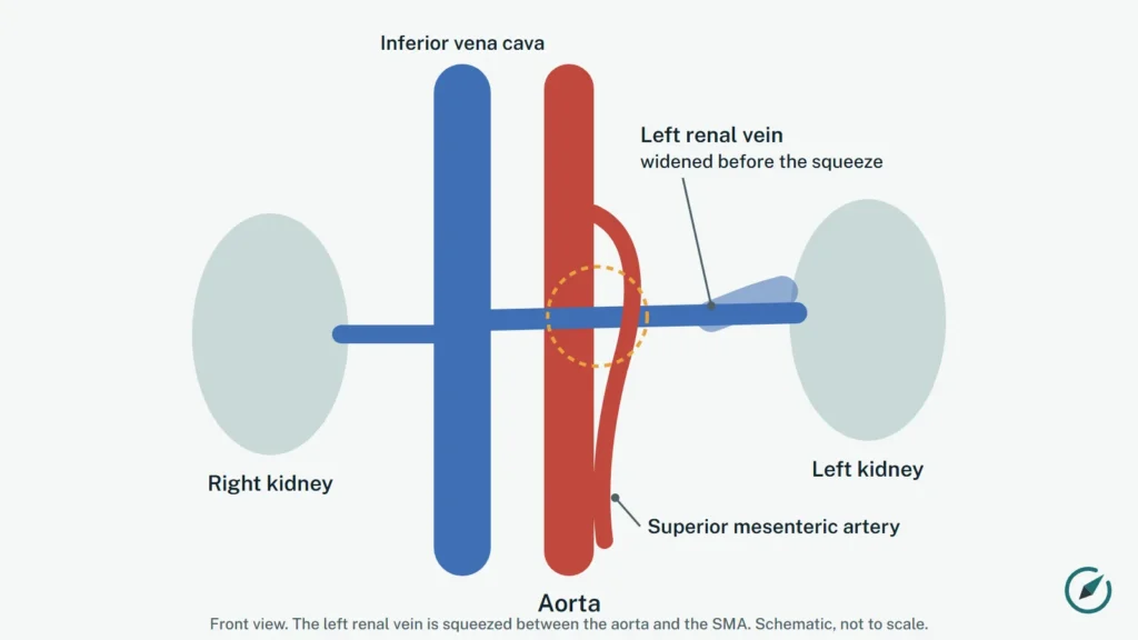
Nutcracker syndrome is a condition in which the left renal vein, the vein that drains blood from the left kidney, is squeezed between the aorta and the superior mesenteric artery, like a nut in a nutcracker. The squeeze can cause blood in the urine, flank or abdominal pain and other symptoms [1][2].
Key points
- Compression of the left renal vein is common on scans of people without symptoms; it is called nutcracker syndrome only when it causes symptoms [3][4].
- Typical signs include blood or protein in the urine, pain, and dizziness when standing up [2].
- Diagnosis usually combines Doppler ultrasound, CT or MRI and sometimes pressure measurements inside the veins [2][1].
- Children often outgrow it, so watchful waiting is common; adults more often need a procedure [2].
- An international expert panel agreed that imaging is required to confirm it, but not on exact cut-off values [3].
What happens in the body
The left renal vein crosses in front of the aorta, just below the point where the superior mesenteric artery branches off. If the space between the two arteries is narrow, the vein is compressed and blood backs up toward the kidney. Over time blood may find other routes, for example through the ovarian or testicular vein and the veins of the pelvis [2][5].
In a less common form, called posterior nutcracker syndrome, a left renal vein that runs behind the aorta is squeezed between the aorta and the spine [5].
How nutcracker syndrome squeezes the kidney vein (4 steps)

1. Normal anatomy. The left renal vein carries blood from the left kidney to the inferior vena cava. On the way it crosses in front of the aorta, passing under the superior mesenteric artery (SMA). Source: study 1, study 2, study 3, study 4.

2. The nutcracker squeeze. In nutcracker syndrome the vein is pinched between the SMA in front and the aorta behind, most often because the angle between these two arteries is narrow. The part of the vein on the kidney side of the squeeze becomes widened. Source: study 1, study 2, study 3, study 4.

3. Blood takes other routes. Pressure builds up in the kidney vein. Blood can flow backward into the left ovarian or testicular vein and on to pelvic veins. The congestion can cause blood in the urine and flank pain; in a 250-patient study, 48% had blood in the urine. Source: study 1, study 2, study 3, study 4, study 5.

4. One treatment: vein transposition. In left renal vein transposition, surgeons cut the vein off the vena cava and sew it back on lower down, away from the squeeze. A review of 24 studies found symptom resolution in 92% after this operation, but 28.5% needed another procedure. Source: study 1, study 2, study 3, study 4.
Prefer a video? Watch the 90-second captioned video explainer.
Nutcracker phenomenon or nutcracker syndrome?
When the vein is compressed but there are no symptoms, doctors call it the nutcracker phenomenon; nutcracker syndrome means the compression causes a recognizable set of symptoms [4]. The difference matters, because signs of compression are frequent in healthy people. In a study of CT scans of 324 healthy kidney donors, one sign of compression (a narrow angle under 41 degrees between the aorta and the SMA) was found in 30.5% of them, and the authors called for better criteria [6].
Symptoms
Reported symptoms include [2]:
- blood in the urine (visible or found on a test);
- protein in the urine;
- pain in the left flank or abdomen;
- dizziness or fainting when standing up (orthostatic intolerance);
- pelvic pain and enlarged pelvic veins, which overlap with pelvic congestion syndrome.
Long-standing high pressure in the vein carries a risk of kidney damage and of a blood clot in the renal vein [1].
How it is diagnosed
Doctors usually work step by step: history and examination, then Doppler ultrasound, CT or MRI, and in selected cases a venogram with intravascular ultrasound and measurement of the pressure difference between the renal vein and the main vein of the body (the renocaval pressure gradient) [2][1]. The international Delphi consensus of 20 venous experts agreed that imaging is needed to confirm the diagnosis, but the experts did not agree on specific cut-off values [3].
Ultrasound for nutcracker syndrome
Doppler ultrasound, a painless scan that also shows how fast blood flows, is often the first imaging test when nutcracker syndrome is suspected [9]. A systematic review that proposed a step-by-step plan advised duplex ultrasound for every patient, with CT or MRI added when needed [12].
The scan compares the left renal vein at the squeeze, between the aorta and the SMA, with the wider part of the vein closer to the kidney [9]:
- Diameter ratio: the width of the vein near the kidney divided by its width at the squeeze. A ratio above 4 has been described in nutcracker syndrome.
- Peak velocity ratio: the top blood speed at the narrowing divided by the speed before it. A ratio above 5 has been described.
In one study of 52 people with blood in the urine, compared with CT, a velocity ratio measured standing up picked up 96.4% of cases (sensitivity) and correctly cleared 79.2% of people without the problem (specificity); it worked better than the diameter ratio [10]. Results can change with body position and breathing [13], technical settings such as the Doppler angle matter [11], and accurate results are described in expert hands [8]. If a procedure is being considered, the same review advised a venogram with measurement of the renocaval pressure gradient and, where available, intravascular ultrasound [12].
Treatment
Treatment depends on how severe the symptoms are [1].
- Watchful waiting: conservative management is appropriate especially in children, who tend to outgrow the condition. Medicines of the ACE inhibitor type have been used for orthostatic symptoms in children [2].
- Surgery: moving the left renal vein to a new position on the main vein (left renal vein transposition) is described as the standard operation, and the expert consensus named it the first-choice operation [2][3]. Kidney autotransplantation is reserved for difficult cases [2].
- Stents: a stent inside the vein is less invasive, but the expert panel agreed that the risk of the stent moving outweighs its advantages [3]. A stent placed around the outside of the vein by keyhole surgery is a newer option [2].
Nutcracker syndrome, SMA syndrome and MALS together
The same narrow space between the aorta and the SMA can compress both the left renal vein (nutcracker syndrome) and the duodenum (SMA syndrome). Doctors have reported a patient with nutcracker syndrome and possible MALS together, and another patient with MALS, SMA syndrome and nutcracker syndrome at once [7][8]. These combinations are rare but explain why some people have symptoms that do not fit a single diagnosis. See also What is MALS.
How well does treatment work? See treatment results from every published study: symptom relief, blood in the urine, stent migration and repeat procedures, side by side.
Read more: left renal vein transposition and other nutcracker operations and nutcracker syndrome, varicocele and pelvic veins in men.
Where is it treated? See US hospitals that publish on nutcracker syndrome, by state.
ICD-10 code
The US ICD-10-CM 2026 code set has no specific entry for nutcracker syndrome. A general code for compression of a vein, I87.1, exists, and the code your doctor uses may depend on the main symptom.
Questions to ask your doctor
- Is this the nutcracker phenomenon seen on a scan, or do my symptoms fit nutcracker syndrome?
- Which tests confirmed it, and was the pressure in the vein measured?
- Could other causes of blood in my urine or my pain have been missed?
- What are the risks and benefits of waiting compared with surgery or a stent?
Current clinical studies
3 registered studies, 1 open or opening soon.
| Study | Status | Type / phase | Where | Last update |
|---|---|---|---|---|
| Lumbar Vein Embolization for Chronic Headaches With Nutcracker Physiology |
Recruiting | Interventional | United States | 2025-11-18 |
| Body Composition, Urine Protein, and Kidney Blood Flow After Nutritional Counseling in Children With Nutcracker Syndrome |
Completed | Observational | Turkey (Türkiye) | 2026-10-09 |
| Renal Autotransplantation; Case Series |
Status unknown | Observational | United States | 2024-03-27 |
Source: ClinicalTrials.gov (U.S. National Library of Medicine), checked November 17, 2026. Status is shown as reported by the registry. A listing is not an endorsement and does not mean a study is right for you: read the official record and discuss it with your doctor.
See all conditions on our clinical trials tracker.
Sources
- Ananthan K, Onida S, Davies AH. Nutcracker Syndrome: An Update on Current Diagnostic Criteria and Management Guidelines. Eur J Vasc Endovasc Surg. 2017;53(6):886-894. PubMed 28356209
- Kolber MK, Cui Z, Chen CK, et al. Nutcracker syndrome: diagnosis and therapy. Cardiovasc Diagn Ther. 2021;11(5):1140-1149. PubMed 34815965
- Heilijgers F, Gloviczki P, O'Sullivan G, et al. Nutcracker syndrome (a Delphi consensus). J Vasc Surg Venous Lymphat Disord. 2025;13(1):101970. PubMed 39362632
- Granata A, Distefano G, Sturiale A, et al. From Nutcracker Phenomenon to Nutcracker Syndrome: A Pictorial Review. Diagnostics (Basel). 2021;11(1). PubMed 33440614
- Said SM, Gloviczki P, Kalra M, et al. Renal nutcracker syndrome: surgical options. Semin Vasc Surg. 2013;26(1):35-42. PubMed 23932560
- Ribeiro FS, Puech-Leao P, Zerati AE, et al. Prevalence of left renal vein compression (nutcracker phenomenon) signs on computed tomography angiography of healthy individuals. J Vasc Surg Venous Lymphat Disord. 2020;8(6):1058-1065. PubMed 32335330
- Donnelly L, Turner B, Davies AH. Atypical case of coexistent vascular compression syndromes: median arcuate ligament syndrome and nutcracker syndrome. BMJ Case Rep. 2023;16(11). PubMed 37989330
- Farina R, Gozzo C, Foti PV, et al. A man with the rare simultaneous combination of three abdominal vascular compression syndromes: median arcuate ligament syndrome, superior mesenteric artery syndrome, and nutcracker syndrome. Radiol Case Rep. 2021;16(6):1264-1270. PubMed 33854661
- Kubiszewski K, Clingan J, Caserta MP. Sonographic Hallmarks of Nutcracker Syndrome. Ultrasound Q. 2026;42(1). PubMed 41705917
- Romera-Villegas A, Vila-Coll R, Cairols-Castellote M, et al. The importance of a standing position in the diagnosis of Nutcracker phenomenon by duplex sonography. Int Angiol. 2009;28(6):461-8. PubMed 20087283
- Kim SH. Doppler US and CT Diagnosis of Nutcracker Syndrome. Korean J Radiol. 2019;20(12):1627-1637. PubMed 31854150
- Nastasi DR, Fraser AR, Williams AB, et al. A systematic review on nutcracker syndrome and proposed diagnostic algorithm. J Vasc Surg Venous Lymphat Disord. 2022;10(6):1410-1416. PubMed 36007798
- Kim SH. The role of Doppler ultrasonography in the detection and management of nutcracker syndrome. Ultrasonography. 2025;44(1):31-41. PubMed 39710849
MALS Compass is an independent educational website, not a medical provider, and is not affiliated with any hospital, clinic or university. This page is educational information and does not replace advice from your own doctor. For any question about your health, please contact your doctor or an official healthcare institution.