Celiac artery anatomy and the median arcuate ligament
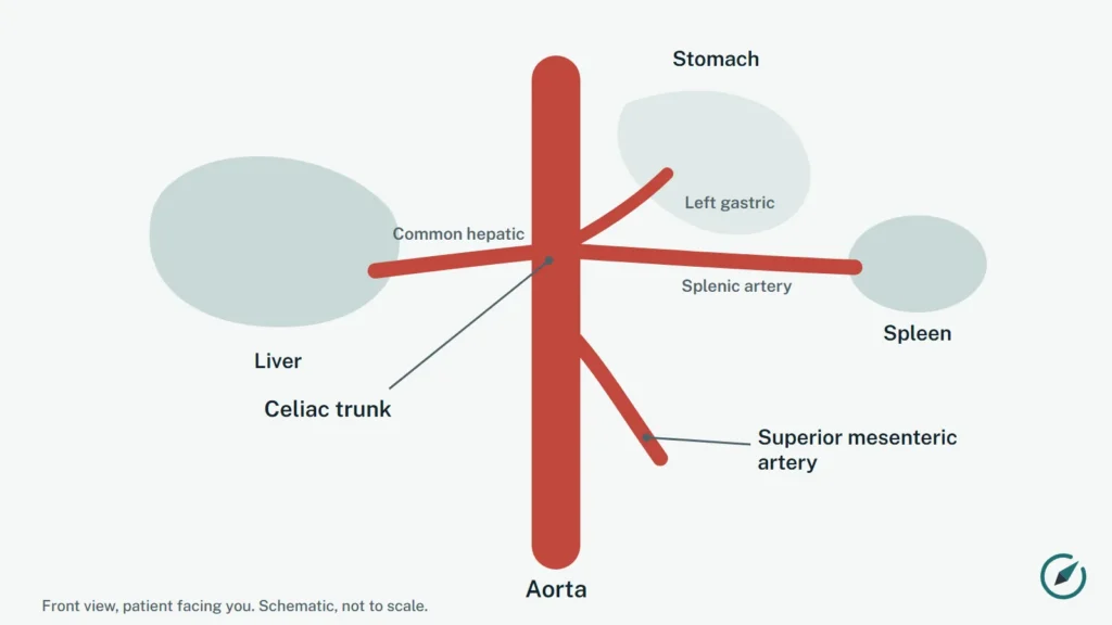
The celiac artery (also called the celiac trunk) is an artery of the upper abdomen that leaves the aorta and splits into three main branches: the left gastric artery, the splenic artery and the common hepatic artery [1]. Understanding this anatomy helps explain how a nearby ligament can press on the artery in median arcuate ligament syndrome (MALS).
Key points
- The celiac artery usually divides into the left gastric, splenic and common hepatic arteries [1].
- Variations in this branching pattern are common: about 1 in 10 people have an incomplete celiac trunk [1].
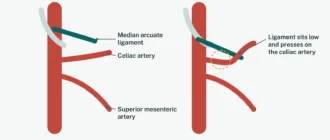
- The median arcuate ligament is a fibrous arch of the diaphragm that usually passes above the start of the celiac artery [9].
- Compression can happen when the artery starts unusually high, when the ligament or diaphragm muscle sits unusually low, or when nerve tissue around the artery is bulky [4][9].
- The normal celiac artery curves downward and then upward, so a "hook" shape on a scan is not proof of compression by itself [5].
Where the celiac artery is
The aorta runs down through an opening in the diaphragm (the muscle that separates the chest from the abdomen). Anatomy textbooks describe the celiac artery as the first branch that leaves the front of the aorta just below this opening, with the superior mesenteric artery about 1 cm lower; in a study of 99 cadavers, however, the outer distance between the two ranged from 0 to 20 mm, and in 57.6% they lay directly next to each other [10]. These arteries are connected by smaller collateral vessels [11]; when the celiac artery is compressed, the changed blood flow has been linked to aneurysms of the celiac artery or of these collaterals [7].
Normal branches and common variations
In a study that combined 43 cadavers and 596 CT angiography scans, 90.5% of people had a complete celiac trunk giving off all three main branches, and 9.5% had an incomplete trunk where one of the branches came off elsewhere [1]. A separate CT study of 340 people found the classic pattern of celiac and hepatic arteries in 70.6%, with the rest showing one of several variant patterns [2]. Surgeons and radiologists need to know about these variations before operations or procedures in the upper abdomen [2][6].
The median arcuate ligament
On either side of the opening for the aorta, the diaphragm has two muscular bands called the crura. They are joined by a fibrous arch: the median arcuate ligament [9]. In most people the ligament passes above the start of the celiac artery; in some it inserts lower and crosses the first part of the artery [9].
A review of the anatomy of celiac artery compression described several ways the artery can be pinched [4]:
- the celiac artery leaves the aorta higher than usual (a "high take-off") and runs under a normally placed ligament;
- the artery starts in the usual place but the crura and ligament are longer than usual;
- a large celiac ganglion (a cluster of nerves around the artery) presses on it;
- a combination of these.
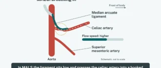
The degree of compression can change with breathing [12]: in one ultrasound study of 10 people with proven MALS, the median blood flow speed in the celiac artery was higher when holding the breath out (287 cm/s) than when holding it in (239 cm/s) [8]. Read more about how this is tested on our page about how MALS is diagnosed.
What a normal artery looks like on a scan
A study of CT angiography scans in 321 healthy kidney donors measured the angle of the celiac artery. In all of them it left the aorta at less than 90 degrees, and in 95% it then turned upward before its first branch [5]. The authors concluded that this natural downward-then-upward course, sometimes described as a hook or J shape, should not be read as a sign of compression on its own. In the same group, 3.4% had a narrowing of more than 50% at the start of the artery without any symptoms [5].
This is one reason why a radiology report that mentions a narrowed or hooked celiac artery does not by itself mean that someone has MALS. Doctors look at the whole picture: symptoms, other possible causes, and how the narrowing behaves; there is no agreed set of diagnostic criteria for MALS [3].
Why the anatomy matters for patients
- Diagnosis: CT angiography shows the course of the artery and its branches without a catheter [6].
- Other conditions: a narrowed celiac artery can also be caused by hardening of the arteries [11]; see celiac artery stenosis.
- Surgery: knowing the branching pattern helps the surgical team plan any operation in the area [2].
Questions to ask your doctor
- Does my scan show a normal celiac artery branching pattern or a variation?
- Was the narrowing measured on images taken when breathing in and when breathing out?
- Could the shape of my artery be normal for me, rather than a sign of compression?
Sources
- Marco-Clement I, Martinez-Barco A, Ahumada N, et al. Anatomical variations of the celiac trunk: cadaveric and radiological study. Surg Radiol Anat. 2016;38(4):501-10. PubMed 26267305
- Acar G, Cicekcibasi AE, Koplay M, et al. Anatomical Variations of the Celiac Trunk and Hepatic Arteries with a New Classification, Based on CT Angiography. J Coll Physicians Surg Pak. 2022;32(1):9-14. PubMed 34983140
- Goodall R, Langridge B, Onida S, et al. Median arcuate ligament syndrome. J Vasc Surg. 2020;71(6):2170-2176. PubMed 31882314
- Loukas M, Pinyard J, Vaid S, et al. Clinical anatomy of celiac artery compression syndrome: a review. Clin Anat. 2007;20(6):612-7. PubMed 17309066
- Petnys A, Puech-Leao P, Zerati AE, et al. Prevalence of signs of celiac axis compression by the median arcuate ligament on computed tomography angiography in asymptomatic patients. J Vasc Surg. 2018;68(6):1782-1787. PubMed 29914831
- Kumar P, Singh A, Triveni GS, et al. Celiac trunk arterial variations and their clinical implications: Role of imaging. Indian J Gastroenterol. 2024;43(6):1099-1110. PubMed 39243342
- Tracci MC. Median arcuate ligament compression of the mesenteric vasculature. Tech Vasc Interv Radiol. 2015;18(1):43-50. PubMed 25814203
- Romer C, Fischer T, Haase O, et al. Assessment of celiac artery compression using color-coded duplex sonography. Clin Hemorheol Microcirc. 2020;76(3):413-423. PubMed 32675404
- Horton KM, Talamini MA, Fishman EK. Median arcuate ligament syndrome: evaluation with CT angiography. Radiographics. 2005;25(5):1177-82. PubMed 16160104
- Katz-Summercorn A, Bridger J. A cadaveric study of the anatomical variation of the origins of the celiac trunk and the superior mesenteric artery: a role in median arcuate ligament syndrome? Clin Anat. 2013;26(8):971-4. PubMed 23554065
- Park CM, Chung JW, Kim HB, et al. Celiac axis stenosis: incidence and etiologies in asymptomatic individuals. Korean J Radiol. 2001;2(1):8-13. PubMed 11752963
- Wang XM, Hua XP, Zheng GL. Celiac Artery Compression Syndrome Evaluated with 3-D Contrast-Enhanced Ultrasonography: a New Approach. Ultrasound Med Biol. 2018;44(1):243-250. PubMed 29079396
MALS Compass is an independent educational website, not a medical provider, and is not affiliated with any hospital, clinic or university. This page is educational information and does not replace advice from your own doctor. For any question about your health, please contact your doctor or an official healthcare institution.