 Exploring the Potential of Ultrasound in Diagnosing Infertility: A Promising Approach
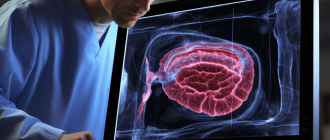
Infertility affects millions of couples worldwide, causing emotional distress and frustration as they struggle to conceive a child. Fortunately, advancements in medical technology have paved the way for more accurate and efficient diagnoses. One such tool is ultrasound, a non-invasive imaging technique that uses sound waves to create images of the reproductive organs.
Ultrasound has become an invaluable tool in the field of infertility diagnoses. It allows doctors to visualize the uterus, fallopian tubes, and ovaries, providing important information about their structure and function. This can help identify potential issues such as fibroids, polyps, or ovarian cysts that may be interfering with fertility.
Additionally, ultrasound can be used to monitor the growth and development of follicles in the ovaries, which play a crucial role in ovulation. By tracking the size and number of follicles, doctors can determine the optimal time for fertility treatments such as in vitro fertilization (IVF) or intrauterine insemination (IUI).
Furthermore, ultrasound-guided procedures such as hysterosalpingography (HSG) or sonohysterography (SHG) can be performed to evaluate the fallopian tubes and the uterine cavity. These procedures can help identify blockages or abnormalities that may be hindering conception.
Overall, ultrasound is a valuable tool in diagnosing infertility. It provides vital information about the reproductive organs and helps doctors tailor treatment plans to each individual’s specific needs. With the help of ultrasound, couples struggling with infertility can have a better understanding of their reproductive health and take steps towards achieving their dream of starting a family.
Ovarian Assessment
Ovarian assessment is an essential part of infertility diagnosis. Ultrasound technology plays a significant role in evaluating the condition of the ovaries. It allows doctors to visualize the size, shape, and structure of the ovaries, as well as monitor the growth and development of follicles.
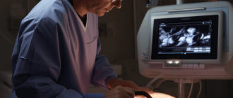
During an ovarian assessment, the ultrasound probe is placed on the abdomen or inserted into the vagina to obtain clear images of the ovaries. The procedure is non-invasive and painless, making it a preferred method for evaluating ovarian health.
Ultrasound can help identify various ovarian conditions that may contribute to infertility, such as polycystic ovary syndrome (PCOS), ovarian cysts, and ovarian tumors. It can also assess the ovarian reserve, which refers to the quantity and quality of eggs remaining in the ovaries.
By examining the ovaries using ultrasound, doctors can determine the number of antral follicles present, which provides an indication of the ovarian reserve. This information is crucial in determining a woman’s fertility potential and guiding treatment options.
Furthermore, ultrasound can aid in monitoring the response of the ovaries to fertility medications during assisted reproductive techniques like in vitro fertilization (IVF). It allows doctors to track follicle growth and determine the optimal time for egg retrieval.
Overall, ovarian assessment using ultrasound is a valuable tool in diagnosing and managing infertility. It provides important information about ovarian health, helps identify potential issues, and guides appropriate treatment strategies to improve fertility outcomes.
Follicle monitoring
Follicle monitoring is a crucial aspect of infertility diagnoses and treatment. Ultrasound technology plays a vital role in monitoring the growth and development of ovarian follicles in women.
Ovarian follicles are small fluid-filled sacs that contain an immature egg. These follicles are responsible for producing and releasing eggs during the menstrual cycle. By monitoring the size and number of follicles, healthcare providers can assess a woman’s ovarian function and determine the best course of action for fertility treatment.
During follicle monitoring, ultrasound imaging is used to visualize the ovaries and measure the size of the follicles. This is typically done using transvaginal ultrasound, where a probe is inserted into the vagina to obtain clear images of the ovaries.
The ultrasound images allow healthcare providers to identify and measure the size of the developing follicles. They can also assess the thickness and appearance of the endometrium, which is the lining of the uterus. This information helps determine the optimal timing for procedures such as intrauterine insemination (IUI) or in vitro fertilization (IVF).
Follicle monitoring is typically performed at regular intervals throughout a woman’s menstrual cycle. This allows healthcare providers to track the growth and size of the follicles over time and make adjustments to the treatment plan if necessary.
In addition to monitoring follicle growth, ultrasound technology can also be used to detect any abnormalities or conditions that may affect fertility, such as ovarian cysts or polycystic ovary syndrome (PCOS).
Overall, follicle monitoring using ultrasound is a non-invasive and effective method for assessing ovarian function and guiding fertility treatment. It provides valuable information that helps healthcare providers tailor treatment plans to each individual’s needs, increasing the chances of a successful pregnancy.
Ovarian reserve testing
Ovarian reserve testing is a crucial component of infertility diagnoses. It helps determine a woman’s fertility potential and the quantity and quality of her eggs. There are several tests that can be performed to assess ovarian reserve:
- Follicle-stimulating hormone (FSH) level: This blood test measures the amount of FSH, a hormone that stimulates the growth of ovarian follicles. Higher FSH levels may indicate reduced ovarian reserve.
- Estradiol level: Estradiol is a form of estrogen produced by the ovaries. Elevated estradiol levels may indicate decreased ovarian reserve.
- Anti-Müllerian hormone (AMH) level: AMH is a hormone produced by the follicles in the ovaries. Low levels of AMH may indicate reduced ovarian reserve.
- Antral follicle count (AFC): This ultrasound-based test counts the number of small follicles in the ovaries. A lower AFC may suggest diminished ovarian reserve.
- Ovarian volume: Ultrasound imaging can measure the size of the ovaries. Smaller ovarian volumes may be indicative of reduced ovarian reserve.
By combining the results of these tests, healthcare professionals can gain a comprehensive understanding of a woman’s ovarian reserve. This information can help guide fertility treatment decisions and provide insight into the likelihood of success with certain interventions.
Polycystic ovarian syndrome diagnosis
Polycystic ovarian syndrome (PCOS) is a common hormonal disorder that affects women of reproductive age. It is characterized by the presence of multiple cysts on the ovaries, irregular menstrual cycles, and high levels of male hormones.
Ultrasound imaging is a valuable tool for diagnosing PCOS. Transvaginal ultrasound is often used to visualize the ovaries and detect the presence of cysts. The ultrasound images can show enlarged ovaries with multiple small cysts arranged around the periphery, giving them a “string of pearls” appearance.
In addition to visualizing the cysts, ultrasound can also help evaluate the thickness of the endometrium and the presence of any uterine abnormalities. These findings can help determine the cause of infertility in women with PCOS.
It is important to note that while ultrasound can provide valuable information for diagnosing PCOS, it is not the only diagnostic tool. Other tests, such as hormonal blood tests and physical exams, may also be necessary to confirm the diagnosis.
If a woman is diagnosed with PCOS, ultrasound can also be used to monitor the response to treatment. Regular ultrasound examinations can help assess the size of the cysts and the overall health of the ovaries, allowing for adjustments to the treatment plan if necessary.
Ultrasound imaging plays a crucial role in the diagnosis and management of polycystic ovarian syndrome. It allows for the visualization of ovarian cysts and helps determine the cause of infertility in women with PCOS. Regular ultrasound examinations can also monitor the response to treatment and ensure the overall health of the ovaries.
Uterine Evaluation
Ultrasound is a valuable tool for evaluating the uterus in cases of infertility. It allows for a detailed examination of the uterus, including its size, shape, and position.
One common cause of infertility is uterine abnormalities, such as fibroids or polyps. These can be easily detected using ultrasound, which can provide a clear image of any abnormalities in the uterine lining.
In addition to detecting abnormalities, ultrasound can also be used to evaluate the thickness of the uterine lining. This is important because a thin lining can make it difficult for a fertilized egg to implant and grow.
During a uterine evaluation, the ultrasound probe is inserted into the vagina to obtain a clear image of the uterus. The probe emits sound waves that bounce off the uterine structures and create an image on a monitor.
In some cases, a saline infusion sonogram may be performed to provide a more detailed evaluation of the uterine lining. This involves injecting saline into the uterus and then performing an ultrasound to examine the lining.
Overall, ultrasound is an essential tool for evaluating the uterus in cases of infertility. It allows for the detection of abnormalities and provides valuable information about the uterine lining, helping to guide treatment decisions and improve the chances of pregnancy.
Uterine fibroids
Uterine fibroids are non-cancerous growths that develop in the uterus. They are also known as leiomyomas or myomas. Fibroids can vary in size, ranging from small seedlings to large masses that can distort the shape of the uterus.
These growths are quite common, with many women developing fibroids at some point in their lives. While the exact cause of uterine fibroids is unknown, hormonal imbalances and genetic factors are believed to play a role in their development.
Symptoms of uterine fibroids can vary depending on their size and location. Some women may experience heavy or prolonged menstrual periods, pelvic pain, frequent urination, or difficulty getting pregnant. In some cases, fibroids may not cause any noticeable symptoms and go undetected until a routine pelvic examination or ultrasound is performed.
Ultrasound imaging is commonly used to diagnose uterine fibroids. During an ultrasound examination, sound waves are used to create images of the uterus and any fibroids present. This non-invasive imaging technique allows healthcare providers to determine the size, location, and characteristics of the fibroids.
Once uterine fibroids are diagnosed, treatment options can be discussed with the patient. In some cases, no treatment may be necessary if the fibroids are small and not causing any symptoms. However, if fibroids are causing pain or fertility issues, treatment options may include medication, minimally invasive procedures, or surgery.
Uterine fibroids are common growths that can develop in the uterus. They can range in size and may cause symptoms such as heavy periods or pelvic pain. Ultrasound imaging is a valuable tool for diagnosing and evaluating uterine fibroids, allowing for appropriate treatment options to be considered.
Uterine abnormalities
Ultrasound is a valuable tool for diagnosing uterine abnormalities that can contribute to infertility. These abnormalities can include structural issues such as fibroids, polyps, or septum, as well as functional abnormalities such as endometrial hyperplasia or adenomyosis.
Using ultrasound, the size, shape, and position of the uterus can be visualized. Fibroids, which are non-cancerous growths in the uterus, can be detected and characterized using ultrasound. The location and size of fibroids can affect fertility and may require treatment to improve the chances of conception.
Polyps, which are small growths on the lining of the uterus, can also be visualized using ultrasound. These polyps can interfere with implantation and may need to be removed to improve fertility outcomes.
A septum, which is a band of tissue dividing the uterus, can also be identified using ultrasound. This structural abnormality can affect the implantation of an embryo and may require surgical correction.
Functional abnormalities such as endometrial hyperplasia, which is an overgrowth of the uterine lining, or adenomyosis, which is the presence of endometrial tissue within the muscle of the uterus, can also be diagnosed using ultrasound. These conditions can affect fertility and may require treatment to improve the chances of conception.
| Uterine abnormality | Ultrasound findings | Treatment options |
| Fibroids | Enlarged uterus, hypoechoic masses | Medication, surgery |
| Polyps | Hyperechoic masses on the uterine lining | Polypectomy |
| Septum | Band of tissue dividing the uterus | Surgical correction |
| Endometrial hyperplasia | Thickened uterine lining | Medication, surgery |
| Adenomyosis | Enlarged uterus with heterogenous texture | Medication, surgery |
Ultrasound plays a crucial role in diagnosing uterine abnormalities that can contribute to infertility. By visualizing the uterus and identifying any structural or functional issues, ultrasound helps guide appropriate treatment options to improve fertility outcomes.
Endometrial lining
The endometrial lining plays a crucial role in the reproductive process as it provides the necessary environment for implantation and pregnancy. Ultrasound imaging is a valuable tool in evaluating the endometrial lining and assessing its thickness and texture.
During the menstrual cycle, the endometrial lining undergoes changes under the influence of hormones. In the early phase of the cycle, the lining is thin and appears as a single layer on ultrasound. As the cycle progresses and ovulation approaches, the endometrium thickens and becomes more vascularized.
Ultrasound can help determine the optimal time for conception by monitoring the changes in the endometrial lining. A thick, well-developed lining with a trilaminar appearance indicates a receptive environment for implantation. On the other hand, a thin or irregularly thickened lining may suggest potential issues with fertility.
In addition to assessing the thickness and texture, ultrasound can also detect abnormalities in the endometrial lining. Polyps, fibroids, and other structural abnormalities can be visualized and evaluated, providing important information for infertility diagnoses.
Overall, ultrasound imaging of the endometrial lining is a non-invasive and effective method for evaluating fertility and diagnosing potential issues. It allows for accurate assessment of the lining’s thickness, texture, and structural abnormalities, helping healthcare providers make informed decisions regarding infertility treatment.
Tubal Testing
Tubal testing is an important part of diagnosing infertility in women. The fallopian tubes play a crucial role in the reproductive process, as they are responsible for carrying the eggs from the ovaries to the uterus. If there is a blockage or damage to the fallopian tubes, it can prevent the sperm from reaching the egg or the fertilized egg from reaching the uterus, resulting in infertility.
There are several methods used to test the fallopian tubes for any abnormalities. One common method is a hysterosalpingogram (HSG), which involves injecting a contrast dye into the uterus and taking X-ray images. The dye allows the doctor to visualize the fallopian tubes and check for any blockages or abnormalities. This test can help determine if the fallopian tubes are open and functioning properly.
Another method of tubal testing is a saline infusion sonohysterography (SIS). This procedure involves injecting saline into the uterus and using ultrasound to create images of the fallopian tubes. The saline helps to expand the uterine cavity and allows the doctor to detect any abnormalities or blockages in the fallopian tubes.
In some cases, a laparoscopy may be recommended to directly visualize the fallopian tubes. This minimally invasive procedure involves making small incisions in the abdomen and inserting a laparoscope, which is a thin tube with a camera attached. The doctor can then examine the fallopian tubes and surrounding structures to check for any abnormalities.
Tubal testing is an important step in diagnosing infertility and determining the best course of treatment. By evaluating the condition of the fallopian tubes, doctors can identify any issues that may be causing infertility and recommend appropriate interventions. These testing methods are generally safe and effective in providing valuable information about the health of the fallopian tubes.
HSG vs ultrasound
When it comes to diagnosing infertility, two common imaging techniques are hysterosalpingography (HSG) and ultrasound. Both methods have their advantages and disadvantages, and the choice between them depends on various factors.
HSG is a radiologic procedure that involves injecting a contrast dye into the uterus and fallopian tubes. The dye helps visualize any abnormalities or blockages that may be present in these reproductive organs. HSG is particularly useful for evaluating tubal patency, detecting uterine abnormalities such as fibroids or polyps, and identifying any structural issues that may contribute to infertility.
On the other hand, ultrasound uses high-frequency sound waves to create images of the reproductive organs. It is a non-invasive procedure that does not involve the use of radiation or contrast dye. Ultrasound can provide valuable information about the size and shape of the uterus, the thickness of the endometrium, and the presence of ovarian cysts or tumors. It can also be used to monitor follicle development during fertility treatments.
One advantage of HSG is its ability to provide a more detailed evaluation of the fallopian tubes. It can detect even subtle abnormalities or blockages that may not be visible on ultrasound. HSG is also a one-time procedure that can be performed in the radiology department.
Ultrasound, on the other hand, is a widely available and cost-effective imaging technique. It can be performed in the gynecologist’s office, and multiple sessions can be scheduled to monitor the progress of infertility treatments. Ultrasound is also safer than HSG since it does not involve exposure to radiation or the use of contrast dye.
Both HSG and ultrasound have their own merits and limitations. The choice between them depends on the specific needs of the patient and the information required for the infertility diagnosis. A healthcare provider can help determine the most appropriate imaging technique based on individual circumstances.
Signs of tubal blockage
Tubal blockage refers to the obstruction or blockage of the fallopian tubes, which can prevent the sperm from reaching the egg or the fertilized egg from reaching the uterus. This condition can cause infertility and is often a result of inflammation, infection, or scarring of the fallopian tubes.
There are several signs that may indicate the presence of tubal blockage:
- Pelvic pain: Women with tubal blockage may experience chronic or intermittent pelvic pain, which can range from mild to severe. The pain may be localized or radiate to other areas of the abdomen.
- Irregular menstrual cycles: Tubal blockage can disrupt the normal hormonal balance, leading to irregular periods. Women with tubal blockage may experience longer or shorter menstrual cycles, as well as changes in the flow and duration of their periods.
- Abnormal vaginal discharge: In some cases, tubal blockage can cause an increase in vaginal discharge. The discharge may be thick, cloudy, or have a foul odor.
- Difficulty getting pregnant: Tubal blockage is a common cause of infertility. If you have been trying to conceive for a year or more without success, it is recommended to consult with a healthcare provider to determine if tubal blockage is the underlying cause.
- Painful intercourse: Some women with tubal blockage may experience pain or discomfort during sexual intercourse. This can be due to the inflammation or scarring of the fallopian tubes, which can make intercourse painful.
If you experience any of these signs or symptoms, it is important to seek medical attention. A healthcare provider can perform diagnostic tests, such as ultrasound, to determine if tubal blockage is present and recommend appropriate treatment options.
Monitoring Infertility Treatments
Ultrasound imaging is a valuable tool for monitoring infertility treatments. It allows healthcare professionals to closely monitor the progress of treatments and make necessary adjustments as needed.
During fertility treatments, such as in vitro fertilization (IVF), ultrasounds are used to track the growth and development of the follicles in the ovaries. This helps determine the optimal time for egg retrieval. Ultrasound can also be used to monitor the thickness of the uterine lining, which is crucial for successful embryo implantation.
Transvaginal ultrasound is commonly used during fertility treatments. This involves inserting a small ultrasound probe into the vagina to obtain detailed images of the reproductive organs. It is a safe and non-invasive procedure that provides valuable information about the ovaries, uterus, and fallopian tubes.
Monitoring infertility treatments with ultrasound can help identify any potential issues or complications that may arise. For example, it can detect the presence of ovarian cysts, which can affect fertility. It can also help identify any abnormalities or abnormalities in the uterus or fallopian tubes that may interfere with conception.
Regular ultrasound monitoring throughout infertility treatments allows healthcare professionals to closely track the progress and adjust the treatment plan accordingly. It provides valuable information about the response to medication, the timing of procedures, and the overall success of the treatment. It also gives patients reassurance and peace of mind, knowing that their progress is being closely monitored.
Ultrasound imaging plays a crucial role in monitoring infertility treatments. It allows healthcare professionals to closely monitor the progress of treatments, make necessary adjustments, and identify any potential issues or complications that may arise. It provides valuable information that helps improve the chances of successful conception and pregnancy.
IUI cycle tracking
Tracking the IUI cycle is an essential part of the infertility treatment process. It involves monitoring the ovaries and uterus to determine the optimal time for insemination.
Ultrasound is a commonly used tool for tracking the IUI cycle. It allows healthcare providers to visualize the ovaries and measure the size of the follicles, which contain the eggs. This information helps determine when the eggs are mature and ready for insemination.
During the IUI cycle tracking, ultrasounds are typically performed at regular intervals. The first ultrasound is usually done at the beginning of the menstrual cycle to establish a baseline. Subsequent ultrasounds are then performed to monitor follicle growth and determine the best time for insemination.
Ultrasound can also be used to evaluate the thickness and quality of the uterine lining. A thick and healthy uterine lining is crucial for successful implantation of the fertilized egg.
In addition to ultrasound, other methods may be used for tracking the IUI cycle, such as blood tests to measure hormone levels. These tests can provide further insights into the hormonal changes that occur during the cycle.
By closely monitoring the IUI cycle, healthcare providers can optimize the timing of insemination, increasing the chances of a successful pregnancy. It also allows for adjustments to the treatment plan if necessary, ensuring the best possible outcome for the patients.
IVF cycle monitoring
During an IVF (in vitro fertilization) cycle, ultrasound is used to monitor the progress and development of the egg follicles. This allows fertility specialists to determine the optimal time for egg retrieval.
Early in the IVF cycle, ultrasound imaging is used to assess the baseline condition of the ovaries and uterus. This provides a starting point for comparison throughout the cycle. The ultrasound can also help identify any abnormalities or potential issues that may affect the success of the IVF cycle.
As the cycle progresses, ultrasounds are typically performed every few days to monitor the growth of the follicles. Follicles are small fluid-filled sacs that contain the eggs. The ultrasound allows the specialist to measure the size and number of the follicles, as well as assess the thickness and quality of the uterine lining.
Once the follicles have reached the desired size, a trigger shot is typically administered to stimulate the final maturation of the eggs. Ultrasound is used to confirm the timing of the trigger shot to ensure optimal egg retrieval.
Following the trigger shot, a final ultrasound is performed just before egg retrieval to confirm the readiness of the follicles. This ultrasound helps guide the fertility specialist during the retrieval procedure to ensure all viable eggs are collected.
Overall, ultrasound monitoring plays a crucial role in IVF cycles by providing valuable information about the development of the follicles and the uterine lining. This helps fertility specialists make informed decisions and optimize the chances of a successful IVF cycle.
Early pregnancy confirmation
Ultrasound is commonly used to confirm early pregnancy. It can detect the presence of a gestational sac as early as 4-5 weeks after the last menstrual period. This allows healthcare providers to confirm the pregnancy and estimate the gestational age.
During an early pregnancy ultrasound, the ultrasound technician or doctor will use a transducer, which emits high-frequency sound waves, to create images of the uterus and the developing embryo. The gestational sac, which is the first sign of pregnancy, can be seen as a small fluid-filled structure within the uterus.
If a gestational sac is visualized, the next step is to look for the presence of a fetal pole and heartbeat. The fetal pole is a thickening of tissue within the gestational sac that will eventually develop into the embryo. The presence of a heartbeat confirms that the pregnancy is viable.
Early pregnancy ultrasound can also help identify potential complications, such as ectopic pregnancy or miscarriage. An ectopic pregnancy occurs when the fertilized egg implants outside of the uterus, usually in the fallopian tube. Miscarriage refers to the loss of a pregnancy before 20 weeks.
Overall, early pregnancy confirmation using ultrasound is a safe and effective method. It provides valuable information about the progression of the pregnancy and helps healthcare providers make informed decisions about prenatal care.
Limitations of Ultrasound in Infertility
While ultrasound is a valuable tool in diagnosing infertility, it does have its limitations. It is important for patients and healthcare providers to be aware of these limitations when using ultrasound for infertility diagnoses.
- Cannot identify all causes of infertility: Ultrasound can provide valuable information about the reproductive organs, but it cannot identify all potential causes of infertility. Other diagnostic tools, such as blood tests and genetic testing, may be necessary to fully understand the underlying causes of infertility.
- Dependent on operator skill: The accuracy and effectiveness of ultrasound in diagnosing infertility can vary depending on the skill and experience of the healthcare provider performing the procedure. It is important to choose a healthcare provider who is experienced in performing infertility ultrasound scans.
- Limited visibility: Ultrasound may not be able to provide clear images of certain structures or abnormalities. For example, pelvic adhesions or small ovarian cysts may be difficult to visualize using ultrasound alone. In such cases, additional imaging techniques, such as magnetic resonance imaging (MRI), may be necessary.
- Cannot assess tubal patency: Ultrasound cannot assess the patency or blockage of the fallopian tubes, which is an important factor in female infertility. Other procedures, such as hysterosalpingography or laparoscopy, may be required to evaluate tubal patency.
- Limited in male infertility assessment: While ultrasound can be used to evaluate male reproductive organs, it may not provide a complete assessment of male infertility. Other tests, such as semen analysis, hormone testing, and genetic testing, may be necessary to fully evaluate male infertility.
Despite its limitations, ultrasound remains a valuable tool in diagnosing infertility and providing important information about the reproductive organs. It is often used in conjunction with other diagnostic tools to provide a comprehensive evaluation of infertility cases.
Not a definitive diagnosis
While ultrasound is a valuable tool in diagnosing infertility, it is important to note that it is not always a definitive diagnosis. Ultrasound can provide valuable information about the reproductive organs and identify any visible abnormalities, but it cannot always determine the exact cause of infertility.
There are various factors that can contribute to infertility, and ultrasound may not be able to detect all of them. For example, ultrasound cannot detect issues with egg quality or sperm motility, which are important factors in fertility. Additionally, ultrasound cannot assess the functionality of the fallopian tubes, which play a crucial role in the fertilization process.
It is also important to note that ultrasound results can vary depending on the skill and experience of the sonographer performing the scan. A highly skilled sonographer may be able to detect subtle abnormalities that a less experienced sonographer may miss.
Therefore, while ultrasound can provide valuable information and help guide further diagnostic testing and treatment, it should not be solely relied upon for a definitive infertility diagnosis. It is important to consult with a fertility specialist who can consider the ultrasound results along with other factors and conduct additional tests to determine the underlying cause of infertility.
User-dependent results
When using ultrasound for infertility diagnoses, it is important to note that the results can be user-dependent. This means that the skill and experience of the ultrasound technician can have a significant impact on the accuracy and reliability of the diagnosis.
An ultrasound technician who is well-trained and experienced in infertility diagnoses will be able to accurately identify and interpret the various structures and abnormalities in the reproductive organs. They will know what to look for, how to position the ultrasound probe correctly, and how to adjust the settings to get the best possible image.
On the other hand, an inexperienced or untrained technician may struggle to identify the subtle signs of infertility or may misinterpret the ultrasound images. This can lead to a misdiagnosis or a failure to detect important issues that may be affecting fertility.
Therefore, it is crucial to ensure that ultrasound exams for infertility diagnoses are performed by qualified and experienced technicians. This will help to minimize the risk of user-dependent errors and improve the overall accuracy of the results.
It is also worth noting that user-dependent results can be influenced by external factors such as the quality of the ultrasound machine and the conditions in which the exam is performed. A high-quality machine and a controlled environment can help to optimize the image quality and enhance the accuracy of the results.
While ultrasound is a valuable tool for diagnosing infertility, it is important to consider the user-dependent nature of the results. By ensuring that exams are performed by skilled technicians and in optimal conditions, the accuracy and reliability of the diagnoses can be greatly improved.
When Additional Testing is Needed
If the initial ultrasound results are inconclusive or if further evaluation is required, additional testing may be necessary. The specific tests recommended will depend on the individual circumstances and the suspected cause of infertility.
One common additional test is a transvaginal ultrasound, which involves inserting a small probe into the vagina to obtain more detailed images of the reproductive organs. This test can provide a closer look at the uterus, ovaries, and fallopian tubes, allowing for a more accurate diagnosis.
In some cases, a hysterosalpingogram may be recommended. This is a procedure in which a dye is injected into the uterus and fallopian tubes, and X-rays are taken to evaluate the structure and function of these organs. This test can help identify blockages or abnormalities that may be causing infertility.
Another option is a sonohysterogram, which involves injecting fluid into the uterus and then performing an ultrasound to evaluate the uterine cavity. This test can provide information about the shape and structure of the uterus, and may be helpful in diagnosing conditions such as uterine fibroids or polyps.
In some cases, additional blood tests or hormonal evaluations may be necessary to assess hormone levels and determine if there are any underlying conditions contributing to infertility.
It is important to work closely with a healthcare provider to determine the appropriate additional tests needed and to understand the implications of the results. With the right testing and diagnosis, appropriate treatment options can be explored to help improve fertility outcomes.
Hysteroscopy
Hysteroscopy is a procedure that allows doctors to examine the inside of the uterus using a thin, lighted tube called a hysteroscope. This procedure is commonly used in the diagnosis and treatment of infertility.
During a hysteroscopy, the hysteroscope is inserted through the vagina and cervix and into the uterus. The hysteroscope transmits images of the uterus to a monitor, allowing the doctor to visualize any abnormalities or issues that may be affecting fertility.
There are two types of hysteroscopy: diagnostic hysteroscopy and operative hysteroscopy. Diagnostic hysteroscopy is used to examine the uterus and diagnose any potential causes of infertility, such as polyps, fibroids, or scar tissue. Operative hysteroscopy, on the other hand, allows doctors to treat these conditions by removing polyps, fibroids, or scar tissue, or by performing other necessary procedures to improve fertility.
One of the advantages of hysteroscopy is that it is a minimally invasive procedure. It can be performed on an outpatient basis, meaning that patients can usually go home the same day. Recovery time is typically short, with most women able to resume their normal activities within a day or two.
Hysteroscopy is considered a safe procedure, but as with any medical procedure, there are risks involved. These risks may include infection, bleeding, or damage to the uterus or other organs. It is important for patients to discuss the potential risks and benefits of hysteroscopy with their doctor before undergoing the procedure.
| Advantages of Hysteroscopy | Risks of Hysteroscopy |
| – Minimally invasive | – Infection |
| – Outpatient procedure | – Bleeding |
| – Short recovery time | – Damage to the uterus or other organs |
Laparoscopy
Laparoscopy is a minimally invasive surgical procedure that allows doctors to examine the reproductive organs in the pelvis. It is commonly used to diagnose and treat infertility in both men and women. During the procedure, a small incision is made in the abdomen, and a thin tube with a camera attached to it, called a laparoscope, is inserted. The camera allows the doctor to view the reproductive organs on a monitor.
Laparoscopy can help diagnose conditions such as endometriosis, pelvic inflammatory disease, ovarian cysts, and uterine fibroids, which can contribute to infertility. It can also be used to remove scar tissue, repair fallopian tubes, and treat other abnormalities that may be causing infertility.
One of the main advantages of laparoscopy is that it is less invasive than traditional open surgery. This means that there is less pain and scarring, and a shorter recovery time. Most patients are able to go home on the same day as the procedure and can resume normal activities within a few days.
However, laparoscopy does carry some risks, including infection, bleeding, and damage to surrounding organs. It is important to discuss the potential risks and benefits of the procedure with your doctor before undergoing laparoscopy.
Laparoscopy is a valuable tool in the diagnosis and treatment of infertility. It allows doctors to visualize the reproductive organs and identify any abnormalities that may be contributing to infertility. While it does carry some risks, the benefits of laparoscopy often outweigh the potential complications.
Blood tests
Blood tests are an essential part of diagnosing infertility. They can provide valuable information about hormone levels, ovarian reserve, and overall reproductive health.
One common blood test used for infertility diagnosis is the follicle-stimulating hormone (FSH) test. FSH is produced by the pituitary gland and plays a crucial role in the development and maturation of eggs in the ovaries. High levels of FSH can indicate a decrease in ovarian reserve and potential fertility issues.
Another important blood test is the luteinizing hormone (LH) test. LH is responsible for triggering ovulation and the release of the egg from the ovary. Abnormal levels of LH can indicate problems with ovulation and fertility.
Estradiol is another hormone that is often measured through blood tests. Estradiol is a form of estrogen and is important for the development of the uterine lining. Abnormal levels of estradiol can indicate issues with the menstrual cycle and potential fertility problems.
In addition to hormone levels, blood tests can also check for other factors that may affect fertility, such as thyroid function, insulin resistance, and certain infections.
Overall, blood tests provide valuable insight into a person’s reproductive health and can help identify potential causes of infertility. They are an important tool in the diagnosis and treatment of infertility, allowing healthcare professionals to tailor treatment plans to individual needs.
Q&A:
What is ultrasound and how is it used for infertility diagnoses?
Ultrasound is a medical imaging technique that uses high-frequency sound waves to produce images of the inside of the body. It is commonly used in infertility diagnoses to assess the reproductive organs, such as the uterus, ovaries, and fallopian tubes. Ultrasound can help detect any abnormalities or structural issues that may be causing infertility.
Is ultrasound a painful procedure?
No, ultrasound is a non-invasive and painless procedure. It involves applying a gel to the skin and then moving a transducer over the area being examined. The transducer emits sound waves that bounce back and create images on a screen. Patients may feel slight pressure or discomfort during the procedure, but it is generally well-tolerated.
What are some common fertility issues that ultrasound can detect?
Ultrasound can help detect a variety of fertility issues, including uterine fibroids, polyps, adhesions, ovarian cysts, and structural abnormalities in the reproductive organs. It can also be used to monitor the growth and development of follicles in the ovaries and to track the thickness of the uterine lining. These findings can provide valuable information for diagnosing the cause of infertility.
Can ultrasound help determine the cause of male infertility?
While ultrasound is primarily used to assess the female reproductive organs, it can also be used to evaluate the male reproductive system. Ultrasound can help detect issues such as varicoceles (enlarged veins in the scrotum), testicular tumors, or any structural abnormalities in the testes or prostate. However, additional tests may be needed to fully diagnose the cause of male infertility.
Are there any risks or side effects associated with ultrasound for infertility diagnoses?
Ultrasound is considered a safe and low-risk procedure. It does not involve radiation, and the sound waves used are not harmful. However, in rare cases, patients may experience an allergic reaction to the gel used during the procedure. Additionally, if a transvaginal ultrasound is performed, there is a slight risk of infection. Overall, the benefits of ultrasound in diagnosing infertility outweigh the minimal risks.