 Understanding and Identifying Intestinal Ischemia: A Comprehensive Diagnosis Guide
Intestinal ischemia, also known as mesenteric ischemia, is a condition in which there is a decrease in blood flow to the intestines. This can lead to serious complications, including tissue damage and even death. Prompt diagnosis is crucial in order to initiate appropriate treatment and prevent further harm.
The diagnosis of intestinal ischemia can be challenging, as the symptoms can be nonspecific and vary depending on the severity and location of the ischemia. However, there are several diagnostic tools that can aid in the identification of this condition.
One of the first steps in diagnosing intestinal ischemia is obtaining a thorough medical history and performing a physical examination. The healthcare provider will ask about the patient’s symptoms, medical conditions, and any risk factors for vascular disease. They will also perform a physical examination to look for signs of abdominal pain, distention, or tenderness.
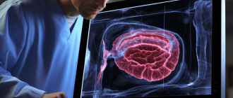
Various imaging studies can also be used to help diagnose intestinal ischemia. These may include abdominal ultrasound, computed tomography (CT) scan, magnetic resonance imaging (MRI), or angiography. These tests can provide detailed images of the blood vessels and help identify any blockages or abnormalities in blood flow.
What tests confirm intestinal ischemia?
Diagnosing intestinal ischemia can be challenging, as its symptoms can be nonspecific and mimic other gastrointestinal conditions. However, several tests can help confirm the presence of intestinal ischemia and determine the extent of the damage.
1. Blood tests: Blood tests can reveal elevated levels of lactate, an indicator of tissue hypoxia, in patients with intestinal ischemia. Other blood markers, such as white blood cell count and C-reactive protein, may also be abnormal.
2. Imaging tests:
- CT angiography: This imaging technique uses a contrast dye and X-rays to visualize the blood vessels in the abdomen. It can help identify blockages or narrowing in the arteries supplying the intestines.
- Mesenteric duplex ultrasound: This non-invasive test uses sound waves to create images of the blood vessels in the abdomen. It can help evaluate blood flow and detect any abnormalities.
- Magnetic resonance angiography (MRA): Similar to CT angiography, MRA uses magnetic fields and radio waves to produce detailed images of the blood vessels. It can provide information about blood flow and detect any blockages or narrowing.
3. Endoscopic tests:
- Colonoscopy: This procedure involves inserting a flexible tube with a camera into the rectum and colon. It can help visualize the lining of the intestines and identify any signs of ischemia.
- Upper endoscopy: Also known as esophagogastroduodenoscopy (EGD), this procedure involves inserting a flexible tube with a camera into the mouth and down the esophagus, stomach, and duodenum. It can help evaluate the upper gastrointestinal tract for signs of ischemia.
4. Exploratory surgery: In some cases, when other tests are inconclusive or urgent intervention is required, exploratory surgery may be performed. This allows direct visualization of the intestines and assessment of blood flow.
It is important to note that the choice of diagnostic tests may vary depending on the individual case and the suspected cause of intestinal ischemia. Healthcare providers will determine the most appropriate tests based on the patient’s symptoms, medical history, and physical examination findings.
What findings on CT angiogram, MRI or catheter angiography confirm ischemia?
When diagnosing intestinal ischemia, imaging techniques such as CT angiogram, MRI, and catheter angiography can provide valuable information to confirm the presence of ischemia. These imaging modalities can help identify specific findings that are indicative of ischemia.
On a CT angiogram, signs of ischemia may include a decreased contrast enhancement of the affected bowel segment, a thickened bowel wall, and the presence of gas within the bowel wall or lumen. These findings suggest compromised blood flow to the intestine, indicating ischemia.
MRI can also be used to evaluate intestinal ischemia and may show similar findings as CT angiogram, such as decreased contrast enhancement and bowel wall thickening. Additionally, MRI can provide information about the blood supply to the intestine and detect any abnormalities or blockages.
Catheter angiography is an invasive procedure that involves injecting contrast dye into the blood vessels to visualize the blood flow. It can be used to directly assess the blood supply to the intestine and identify any blockages or narrowing of the arteries. A lack of blood flow or irregularities in the arterial blood vessels can confirm the presence of ischemia.
Overall, CT angiogram, MRI, and catheter angiography are valuable tools in diagnosing intestinal ischemia. These imaging techniques can provide specific findings that confirm the presence of ischemia, helping guide appropriate treatment decisions.
What blood tests may support the diagnosis?
Blood tests play a crucial role in the diagnosis of intestinal ischemia. Several specific blood markers can help support the diagnosis and provide valuable information about the condition of the intestines.
One of the primary blood tests used is the complete blood count (CBC). This test measures the number of red blood cells, white blood cells, and platelets in the blood. In cases of intestinal ischemia, the CBC may show an increased white blood cell count, indicating inflammation or infection in the intestines.
Another important blood test is the serum lactate level. Intestinal ischemia disrupts blood flow to the intestines, leading to tissue damage and decreased oxygen supply. As a result, the body produces more lactate, which can be measured in the blood. Elevated levels of lactate suggest that there is insufficient oxygen reaching the intestines, supporting the diagnosis of intestinal ischemia.
Additionally, blood tests for markers of inflammation, such as C-reactive protein (CRP) and erythrocyte sedimentation rate (ESR), can be helpful. High levels of these markers indicate inflammation in the body, which can be present in cases of intestinal ischemia.
In some cases, blood tests may also be used to assess organ function and overall health. Liver function tests, kidney function tests, and electrolyte levels can provide valuable information about the patient’s condition and help guide treatment decisions.
Blood tests are essential in supporting the diagnosis of intestinal ischemia. The complete blood count, serum lactate level, markers of inflammation, and organ function tests can provide valuable insights into the condition of the intestines and guide appropriate treatment strategies.
What nonspecific symptoms should raise suspicion for intestinal ischemia?
Intestinal ischemia, also known as mesenteric ischemia, occurs when there is a decrease in blood flow to the intestines. It is a serious condition that can lead to tissue damage and organ failure if not promptly diagnosed and treated. While the symptoms of intestinal ischemia can vary depending on the severity and location of the blockage, there are some nonspecific symptoms that should raise suspicion for this condition.
One of the key nonspecific symptoms of intestinal ischemia is severe abdominal pain. This pain is often described as being constant and crampy in nature, and it may be out of proportion to the physical examination findings. The pain may also worsen after eating and may not be relieved by bowel movements or passing gas.
In addition to abdominal pain, patients with intestinal ischemia may experience other nonspecific symptoms such as nausea and vomiting. These symptoms can be caused by the disruption of normal bowel function and can sometimes be accompanied by a loss of appetite. Patients may also experience weight loss due to a decreased ability to eat and absorb nutrients.
Another nonspecific symptom that may raise suspicion for intestinal ischemia is diarrhea. This can occur due to the inflammation and damage to the intestines caused by reduced blood flow. The diarrhea may be watery and may contain blood or mucus.
Other nonspecific symptoms that may be present in patients with intestinal ischemia include fever, fatigue, and a general feeling of being unwell. These symptoms can be caused by the body’s response to the tissue damage and inflammation that occurs with this condition.
It is important to note that these nonspecific symptoms can also be caused by other conditions, and further diagnostic testing is needed to confirm a diagnosis of intestinal ischemia. However, if a patient presents with these symptoms, it is important for healthcare providers to consider the possibility of intestinal ischemia and to initiate appropriate diagnostic measures to ensure timely treatment.
What is the gold standard for diagnosing intestinal ischemia?
The gold standard for diagnosing intestinal ischemia is mesenteric angiography. This imaging technique uses a contrast dye injected into the blood vessels of the intestines to visualize the blood flow. It provides detailed images of the blood vessels, allowing doctors to identify any blockages or narrowing that may be causing the ischemia. Mesenteric angiography is considered the most accurate method for diagnosing intestinal ischemia and is often used when other tests are inconclusive or to confirm a suspected diagnosis.
Why is angiography considered the gold standard?
Angiography is widely regarded as the gold standard for diagnosing intestinal ischemia due to its accuracy and ability to provide detailed visualization of the blood vessels in the affected area. This diagnostic imaging technique involves the injection of a contrast dye into the blood vessels, followed by X-ray imaging to capture real-time images of the blood flow.
There are several reasons why angiography is considered the preferred method for diagnosing intestinal ischemia:
| Accuracy | Angiography provides highly accurate results, allowing for the precise identification and localization of any blockages or narrowing of the blood vessels in the intestines. This is crucial for determining the extent and severity of the ischemia. |
| Visualization | Angiography offers excellent visualization of the blood vessels, allowing for the detection of even small abnormalities or abnormalities in the vasculature. This is particularly important in cases of intestinal ischemia, where prompt detection and intervention are essential. |
| Real-time imaging | Angiography provides real-time imaging, allowing the physician to closely monitor the blood flow and detect any changes or abnormalities immediately. This can be crucial in guiding treatment decisions and interventions. |
| Therapeutic potential | In addition to its diagnostic value, angiography can also be used for therapeutic purposes. In cases where a blockage or narrowing is identified, the physician can perform minimally invasive procedures, such as angioplasty or stenting, to restore blood flow and alleviate the ischemia. |
Angiography is considered the gold standard for diagnosing intestinal ischemia due to its high accuracy, excellent visualization capabilities, real-time imaging, and therapeutic potential. It plays a crucial role in the timely and accurate diagnosis and management of this condition.
What are its advantages and limitations?
The diagnosis of intestinal ischemia has several advantages, but also some limitations. One of the main advantages is that it allows healthcare professionals to identify this condition early on, which can lead to prompt treatment and improved outcomes for patients. Additionally, the use of diagnostic tests and imaging techniques can provide valuable information about the extent and severity of the ischemia, helping physicians make informed decisions about the most appropriate treatment approach.
Another advantage of diagnosing intestinal ischemia is that it can help differentiate this condition from other causes of abdominal pain and discomfort. This is particularly important as the symptoms of intestinal ischemia can be nonspecific and mimic other gastrointestinal disorders. By accurately diagnosing intestinal ischemia, healthcare professionals can avoid unnecessary procedures or treatments and provide targeted care to the patient.
However, there are also limitations to the diagnosis of intestinal ischemia. One of the main challenges is that the symptoms of intestinal ischemia can be subtle and easily overlooked. This can result in delayed diagnosis and treatment, which can have serious consequences for the patient. Additionally, some diagnostic tests for intestinal ischemia, such as angiography, can be invasive and carry risks of complications.
Furthermore, the accuracy of diagnostic tests for intestinal ischemia can vary. While some tests, such as computed tomography (CT) angiography, can provide highly accurate results, others may be less reliable. This can make the diagnosis of intestinal ischemia challenging, particularly in cases where the symptoms are atypical or the condition is in its early stages.
The diagnosis of intestinal ischemia has several advantages, including early identification and differentiation from other conditions. However, there are also limitations, such as the subtle symptoms and varying accuracy of diagnostic tests. Healthcare professionals must be diligent in considering the possibility of intestinal ischemia and utilizing appropriate diagnostic tools to ensure timely and accurate diagnosis.
When should angiography be performed?
Angiography is a diagnostic test that uses X-ray imaging to visualize blood vessels. In the context of intestinal ischemia, angiography can play a crucial role in determining the extent and location of the ischemic damage.
Angiography should be performed when there is a strong suspicion of intestinal ischemia based on clinical symptoms and other diagnostic tests. Some of the indications for angiography include:
| 1. | Persistent abdominal pain that is not responsive to conservative management. |
| 2. | Unexplained gastrointestinal bleeding. |
| 3. | Signs of peritonitis or sepsis. |
| 4. | Abnormal findings on other imaging modalities, such as CT scan or ultrasound. |
| 5. | High clinical suspicion in patients with risk factors for intestinal ischemia, such as advanced age, cardiovascular disease, or prior abdominal surgery. |
Angiography can provide valuable information about the location and severity of the arterial occlusion, which can guide further management decisions. It can also help identify potential candidates for revascularization procedures, such as angioplasty or bypass surgery.
However, it is important to note that angiography is an invasive procedure and carries some risks, including contrast dye allergy and injury to the blood vessels. Therefore, the decision to perform angiography should be made on a case-by-case basis, weighing the potential benefits against the risks.
Angiography should be considered in patients with suspected intestinal ischemia who meet certain clinical criteria. It can provide important diagnostic information and guide further management decisions in these patients.
What imaging modalities can be used to diagnose intestinal ischemia?
Diagnosing intestinal ischemia can be challenging, as the symptoms can be nonspecific and overlapping with other conditions. However, several imaging modalities can be used to aid in the diagnosis of intestinal ischemia.
1. Abdominal X-ray: An abdominal X-ray can be the initial imaging modality used to evaluate patients with suspected intestinal ischemia. It can help identify signs such as free air, bowel dilation, or air-fluid levels, which may indicate intestinal ischemia.
2. Computed tomography (CT) scan: CT scan is a commonly used imaging modality for diagnosing intestinal ischemia. It provides detailed images of the abdominal organs and blood vessels, allowing for the detection of abnormalities such as bowel wall thickening, pneumatosis intestinalis (gas within the bowel wall), or mesenteric ischemia (reduced blood flow to the intestines).
3. Magnetic resonance imaging (MRI): MRI is another imaging modality that can be used to diagnose intestinal ischemia. It can provide high-resolution images of the abdominal organs and blood vessels, allowing for the visualization of ischemic changes in the intestines.
4. Angiography: Angiography involves the injection of contrast dye into the blood vessels to visualize the blood flow. It can be useful in diagnosing mesenteric ischemia, as it can identify areas of reduced blood flow or blockages in the mesenteric arteries.
5. Doppler ultrasound: Doppler ultrasound uses sound waves to assess blood flow in the arteries and veins. It can be used to evaluate the blood flow in the mesenteric arteries and detect any abnormalities that may indicate intestinal ischemia.
It is important to note that the choice of imaging modality depends on various factors, including the patient’s clinical presentation, availability of resources, and the expertise of the healthcare provider. In some cases, a combination of imaging modalities may be needed to establish a definitive diagnosis of intestinal ischemia.
What is the role of CT angiography?
CT angiography plays a crucial role in the diagnosis of intestinal ischemia. It is a non-invasive imaging technique that uses computed tomography to visualize the blood vessels in the abdomen and pelvis.
By injecting a contrast dye into the patient’s veins, CT angiography allows for the detailed assessment of blood flow and the detection of any abnormalities or blockages in the arteries supplying the intestines. This information is essential in determining the presence and extent of intestinal ischemia.
CT angiography can accurately identify the location and severity of arterial occlusions, thrombosis, embolisms, or other vascular abnormalities that may be causing intestinal ischemia. It is particularly useful in differentiating between different types of ischemia, such as arterial or venous ischemia.
Furthermore, CT angiography provides valuable information about the collateral circulation, which refers to the alternate pathways that blood can take to reach the intestines in case of arterial blockages. This information helps in assessing the potential for revascularization procedures.
Overall, CT angiography is a valuable diagnostic tool in the evaluation of intestinal ischemia. It provides detailed and accurate information about the blood vessels in the abdomen and pelvis, helping clinicians make informed decisions about the appropriate treatment plan for patients with this condition.
What is the role of MRI and MR angiography?
Magnetic Resonance Imaging (MRI) and Magnetic Resonance Angiography (MRA) play an important role in the diagnosis of intestinal ischemia. These imaging techniques are non-invasive and provide detailed information about the blood vessels and the tissues of the intestine.
MRI uses powerful magnets and radio waves to create detailed images of the organs and tissues in the body. It can help identify any abnormalities in the blood vessels and detect signs of ischemia, such as narrowing or blockages.
MRA, on the other hand, focuses specifically on the blood vessels. It uses a contrast dye that is injected into the patient’s veins to help visualize the blood vessels and any abnormalities. MRA can provide detailed images of the blood supply to the intestine, helping to identify any areas of reduced blood flow or blockages.
Together, MRI and MRA can provide valuable information about the extent and location of intestinal ischemia. They can help differentiate between different types of ischemia, such as acute mesenteric ischemia and chronic mesenteric ischemia, and guide further treatment decisions.
In addition to diagnosing intestinal ischemia, MRI and MRA can also be used to monitor the progression of the condition and assess the effectiveness of treatment. Repeat imaging can help determine if the blood flow has improved or if there are any new developments that require intervention.
Overall, MRI and MRA are valuable tools in the diagnosis and management of intestinal ischemia. They provide detailed and non-invasive imaging of the blood vessels and tissues, helping to guide appropriate treatment decisions and monitor the condition over time.
What is the role of ultrasound?
Ultrasound plays a crucial role in the diagnosis of intestinal ischemia. It is a non-invasive imaging technique that uses sound waves to create images of the abdominal organs. Ultrasound can provide valuable information about the blood flow to the intestines, which is essential in diagnosing ischemia.
One of the main advantages of ultrasound is its ability to quickly assess the blood flow in the intestines. Doppler ultrasound, a specialized technique, can measure the speed and direction of blood flow, allowing for the detection of any abnormalities. This is particularly useful in cases of acute intestinal ischemia, where prompt diagnosis is crucial for successful treatment.
In addition to assessing blood flow, ultrasound can also help identify other signs of intestinal ischemia. It can detect thickening of the bowel wall, which may indicate inflammation or ischemic injury. Ultrasound can also identify the presence of gas in the bowel, which can be a sign of bowel obstruction or infarction.
Ultrasound is a safe and cost-effective imaging modality that can be performed at the bedside. It does not involve exposure to ionizing radiation, making it a preferred option for patients who are pregnant or have concerns about radiation exposure. Furthermore, ultrasound can be repeated as needed to monitor the progression of intestinal ischemia or the response to treatment.
Ultrasound plays a crucial role in the diagnosis of intestinal ischemia. It can provide valuable information about blood flow, detect signs of ischemia, and guide further management. Its non-invasive nature and ability to be repeated make it a valuable tool in the evaluation of patients with suspected intestinal ischemia.
What findings on imaging indicate intestinal ischemia?
Imaging plays a crucial role in the diagnosis of intestinal ischemia. Several findings on imaging can indicate the presence of intestinal ischemia, helping physicians make an accurate diagnosis. These findings may include:
- Segmental or diffuse bowel wall thickening
- Decreased or absent bowel wall enhancement
- Presence of pneumatosis intestinalis (gas within the bowel wall)
- Portal venous gas
- Ischemic changes in adjacent organs, such as the liver or spleen
- Ascites (fluid accumulation in the abdominal cavity)
- Abnormalities in the mesenteric vessels, such as thrombosis or embolism
- Bowel dilation
These imaging findings can be identified using various imaging modalities, including computed tomography (CT), magnetic resonance imaging (MRI), and angiography. CT is the most commonly used modality for the diagnosis of intestinal ischemia due to its wide availability and ability to provide detailed images of the bowel.
It is important to note that these imaging findings are not specific to intestinal ischemia and can also be seen in other conditions. Therefore, clinical correlation and a thorough evaluation of the patient’s medical history and symptoms are essential for an accurate diagnosis.
What CT angiography findings suggest intestinal ischemia?
CT angiography is a valuable imaging technique for the diagnosis of intestinal ischemia. It allows for the evaluation of blood flow within the mesenteric vessels and can provide important clues to the presence of ischemia. Several CT angiography findings can suggest the presence of intestinal ischemia:
1. Absence or decreased enhancement of the bowel wall: In cases of intestinal ischemia, the affected bowel segment may show decreased enhancement or even absence of enhancement on CT angiography. This is due to the compromised blood flow to the bowel wall.
2. Presence of bowel wall thickening: Intestinal ischemia can cause edema and inflammation of the bowel wall, leading to thickening. CT angiography can detect this thickening, which may be a sign of ischemia.
3. Presence of bowel dilatation: In some cases of intestinal ischemia, there may be associated bowel dilatation. CT angiography can identify the presence of dilated bowel loops, which may be indicative of ischemia.
4. Presence of mesenteric vessel occlusion or thrombosis: Intestinal ischemia can be caused by occlusion or thrombosis of the mesenteric vessels. CT angiography can visualize these occluded or thrombosed vessels, providing evidence of ischemia.
5. Presence of pneumatosis intestinalis: In severe cases of intestinal ischemia, gas may accumulate within the bowel wall, leading to the presence of pneumatosis intestinalis. CT angiography can detect this gas within the bowel wall, which is a concerning finding for ischemia.
Overall, CT angiography findings suggestive of intestinal ischemia include absence or decreased enhancement of the bowel wall, bowel wall thickening, bowel dilatation, mesenteric vessel occlusion or thrombosis, and the presence of pneumatosis intestinalis. These findings can help guide the diagnosis and management of intestinal ischemia.
What angiography findings confirm reduced blood flow?
Angiography is a valuable diagnostic tool used to assess blood flow in the intestines. It involves the injection of a contrast dye into the blood vessels, which highlights the blood vessels on X-ray images. By analyzing the angiography findings, doctors can confirm if there is reduced blood flow to the intestines.
There are several angiography findings that can confirm reduced blood flow:
| Angiography Finding | Description |
|---|---|
| Filling defects | These are areas where the contrast dye fails to fill the blood vessels, indicating a blockage or narrowing that restricts blood flow. |
| Delayed or absent contrast washout | In normal circumstances, the contrast dye should wash out of the blood vessels relatively quickly. However, in cases of reduced blood flow, the dye may persist for a longer period or fail to wash out completely. |
| Collateral vessel formation | In response to reduced blood flow, the body may form new blood vessels to bypass the blocked or narrowed areas. These collateral vessels can be visualized on angiography and indicate compromised blood flow. |
| Segmental or diffuse narrowing of vessels | Reduced blood flow can cause narrowing of multiple blood vessels in the intestines. This can be seen as segmental or diffuse narrowing on angiography images. |
By identifying these angiography findings, doctors can confirm the presence of reduced blood flow to the intestines and guide further treatment decisions. It is important to note that angiography findings should be interpreted in conjunction with clinical symptoms and other diagnostic tests to make an accurate diagnosis of intestinal ischemia.
How does bowel wall appearance differ in ischemic intestine?
In cases of intestinal ischemia, the appearance of the bowel wall can be significantly different from that of a healthy intestine. Ischemic intestine refers to a condition where there is a lack of blood flow to the intestines, leading to tissue damage and potentially life-threatening complications.
One of the key differences in the appearance of the bowel wall in ischemic intestine is the presence of edema. Edema refers to the accumulation of fluid in the tissues, and in the case of ischemic intestine, it can cause the bowel wall to become thickened and swollen.
In addition to edema, ischemic intestine may also present with hemorrhage. Hemorrhage refers to the escape of blood from blood vessels, and it can manifest as areas of bleeding within the bowel wall. These areas may appear as dark or red patches on imaging studies.
Another characteristic feature of ischemic intestine is the presence of pneumatosis intestinalis. Pneumatosis intestinalis refers to the presence of gas within the bowel wall. This gas can be visualized on imaging studies and appears as small pockets or bubbles within the wall of the intestine.
Furthermore, ischemic intestine may exhibit signs of necrosis. Necrosis refers to tissue death, and in the case of ischemic intestine, it can lead to the formation of black or darkened areas within the bowel wall. These areas are indicative of severe tissue damage and require immediate medical attention.
Overall, the appearance of the bowel wall in ischemic intestine is characterized by edema, hemorrhage, pneumatosis intestinalis, and necrosis. These features can be detected through various imaging modalities, such as computed tomography (CT) scans or ultrasound, and are crucial in diagnosing and managing this potentially life-threatening condition.
How accurate are different diagnostic tests for intestinal ischemia?
Accurate diagnosis of intestinal ischemia is crucial for prompt treatment and improved patient outcomes. There are several diagnostic tests available to assess the presence and severity of intestinal ischemia. The accuracy of these tests varies depending on various factors, including the type of test and the underlying condition of the patient.
One commonly used diagnostic test for intestinal ischemia is abdominal computed tomography (CT) scan. This imaging technique provides detailed images of the abdominal region, allowing doctors to identify any signs of ischemia, such as bowel wall thickening or gas in the bowel wall. The accuracy of CT scan in diagnosing intestinal ischemia ranges from 70% to 95%.
Another diagnostic test is mesenteric angiography, which involves injecting a contrast dye into the blood vessels of the mesentery and taking X-ray images. This test can help identify any blockages or narrowing of the mesenteric arteries, indicating intestinal ischemia. The accuracy of mesenteric angiography is reported to be around 80% to 90%.
Additionally, laboratory tests can be useful in diagnosing intestinal ischemia. Elevated levels of lactate or lactate dehydrogenase in the blood may indicate tissue hypoxia and suggest the presence of ischemia. However, these tests have relatively low specificity and may not be conclusive on their own.
Exploratory laparotomy, a surgical procedure, can also be used to diagnose intestinal ischemia. During this procedure, the surgeon visually inspects the intestines for any signs of ischemia, such as discoloration or necrosis. This method is considered the gold standard for diagnosing intestinal ischemia, with an accuracy of nearly 100%.
The accuracy of different diagnostic tests for intestinal ischemia varies. Abdominal CT scan and mesenteric angiography are effective imaging techniques, while laboratory tests can provide additional information. However, exploratory laparotomy remains the most accurate method for diagnosing intestinal ischemia.
What is the sensitivity of CT angiography?
CT angiography is a widely used imaging technique for the diagnosis of intestinal ischemia. It is a non-invasive procedure that uses a combination of X-rays and computer technology to create detailed images of the blood vessels in the abdomen.
The sensitivity of CT angiography in the diagnosis of intestinal ischemia is relatively high. Studies have shown that it can detect the presence of ischemia with a sensitivity ranging from 80% to 95%. This means that CT angiography has the ability to accurately identify the majority of cases of intestinal ischemia.
CT angiography is particularly useful in detecting acute mesenteric ischemia, which is a severe form of intestinal ischemia that requires immediate medical attention. The high sensitivity of CT angiography allows for early detection of this condition, leading to prompt treatment and improved patient outcomes.
In addition to its high sensitivity, CT angiography also has other advantages. It is a quick and relatively painless procedure that can be performed on an outpatient basis. It does not require the use of contrast agents, which can be beneficial for patients with kidney problems or allergies to contrast materials.
Overall, CT angiography is a valuable diagnostic tool for the detection of intestinal ischemia. Its high sensitivity, along with its other advantages, makes it an important imaging modality in the management of patients with suspected intestinal ischemia.
What is the sensitivity and specificity of angiography?
Angiography is a common diagnostic imaging procedure used to evaluate intestinal ischemia. It involves the injection of contrast dye into the blood vessels of the intestine, followed by X-ray imaging to visualize the blood flow and identify any blockages or abnormalities.
The sensitivity and specificity of angiography in diagnosing intestinal ischemia depend on various factors, including the experience of the radiologist performing the procedure and the quality of the imaging equipment used. However, studies have shown that angiography has a high sensitivity and specificity for detecting intestinal ischemia.
The sensitivity of angiography refers to its ability to correctly identify patients with intestinal ischemia. A high sensitivity means that the test has a low rate of false-negative results, meaning it rarely misses cases of the disease. In the case of angiography, it has been reported to have a sensitivity of over 90% for diagnosing intestinal ischemia.
The specificity of angiography, on the other hand, refers to its ability to correctly identify patients without intestinal ischemia. A high specificity means that the test has a low rate of false-positive results, meaning it rarely misdiagnoses patients without the disease. Angiography has been reported to have a specificity of around 95% for diagnosing intestinal ischemia.
It is important to note that angiography is an invasive procedure and carries certain risks, such as allergic reactions to the contrast dye or damage to the blood vessels. Therefore, it is usually reserved for cases where there is a high suspicion of intestinal ischemia or when other non-invasive imaging tests have been inconclusive.
Angiography is a highly sensitive and specific imaging technique for diagnosing intestinal ischemia. However, its use should be carefully considered, weighing the potential risks and benefits for each individual patient.
How do blood tests and endoscopy compare in accuracy?
When it comes to diagnosing intestinal ischemia, both blood tests and endoscopy play important roles in determining the accuracy of the diagnosis. However, they differ in their approaches and the information they provide.
Blood tests are often the first step in diagnosing intestinal ischemia. These tests measure various markers in the blood that can indicate the presence of ischemia, such as elevated levels of lactate, D-dimer, or white blood cells. While blood tests can provide valuable information, they are not always definitive and may require further investigation.
On the other hand, endoscopy is a more invasive procedure that involves inserting a flexible tube with a camera into the digestive tract. This allows doctors to directly visualize the intestines and identify any signs of ischemia, such as ulcers, bleeding, or narrowing of the blood vessels. Endoscopy can provide more detailed information than blood tests and is often considered more accurate in diagnosing intestinal ischemia.
However, it’s important to note that both blood tests and endoscopy have their limitations. Blood tests can be influenced by other factors, such as dehydration or underlying medical conditions, which may affect their accuracy. Endoscopy, while more accurate, may not always be feasible or readily available in certain situations.
| Comparison | Blood Tests | Endoscopy |
|---|---|---|
| Approach | Non-invasive | Invasive |
| Information provided | Indirect markers of ischemia | Direct visualization of intestines |
| Accuracy | Less definitive, may require further investigation | More accurate in diagnosing intestinal ischemia |
| Limitations | May be influenced by other factors | May not always be feasible or readily available |
In conclusion, both blood tests and endoscopy are important tools in diagnosing intestinal ischemia. While blood tests can provide initial information, endoscopy is often considered more accurate due to its direct visualization of the intestines. However, it’s important for doctors to consider the limitations and availability of these tests when making a diagnosis.
Q&A:
What is intestinal ischemia?
Intestinal ischemia is a condition in which the blood supply to the intestines is reduced or completely blocked, leading to tissue damage and potentially life-threatening complications.
What are the common symptoms of intestinal ischemia?
The common symptoms of intestinal ischemia include abdominal pain, bloody stool, diarrhea, nausea, vomiting, and weight loss. However, the symptoms can vary depending on the severity and location of the ischemia.
How is intestinal ischemia diagnosed?
Intestinal ischemia can be diagnosed through various methods. These include physical examination, blood tests, imaging tests such as CT scan or angiography, and sometimes a colonoscopy or endoscopy to directly visualize the intestines.
What are the risk factors for developing intestinal ischemia?
The risk factors for developing intestinal ischemia include advanced age, atherosclerosis, certain medical conditions such as diabetes or high blood pressure, smoking, and certain medications such as oral contraceptives or vasoconstrictors.
What are the treatment options for intestinal ischemia?
The treatment options for intestinal ischemia depend on the severity and underlying cause of the condition. In some cases, medications to improve blood flow and manage symptoms may be sufficient. However, in more severe cases, surgery may be necessary to remove the damaged portion of the intestine or restore blood flow.
What is intestinal ischemia?
Intestinal ischemia is a condition in which there is a decreased blood flow to the intestines, leading to tissue damage and potential organ failure.
What are the symptoms of intestinal ischemia?
The symptoms of intestinal ischemia can vary depending on the severity of the condition, but common symptoms include abdominal pain, bloody stool, diarrhea, nausea, and vomiting.
How is intestinal ischemia diagnosed?
Intestinal ischemia can be diagnosed through various methods, including physical examination, blood tests, imaging tests such as CT scans or angiography, and sometimes through invasive procedures like endoscopy or laparoscopy.