 Interpreting and Analyzing the Results of an Upper GI Series Test: A Comprehensive Guide
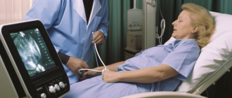
When you experience symptoms such as abdominal pain, difficulty swallowing, or unexplained weight loss, your doctor may order an upper gastrointestinal (GI) series test to help diagnose the cause. This test is a type of X-ray examination that focuses on the upper part of your digestive system, including the esophagus, stomach, and small intestine. Understanding the results of this test can provide valuable information about your digestive health.
During an upper GI series test, you will be asked to drink a contrast material, such as barium, which helps highlight the structures in your digestive system. X-ray images will then be taken as the contrast material moves through your upper GI tract. These images can reveal abnormalities or blockages that may be causing your symptoms.
Once the test is complete, a radiologist will interpret the images and provide a report to your doctor. It’s important to note that the interpretation of the results requires specialized medical knowledge, so it’s best to consult with your doctor for a thorough explanation. However, understanding some common terms and findings can help you have a more informed discussion about your test results.
Overview of Results Analysis
After the upper GI series test, the radiologist will analyze the images to look for any abnormalities or issues in the upper gastrointestinal tract. This analysis will help to determine the cause of symptoms and guide further treatment options.
The radiologist will examine the images for any signs of inflammation, ulcers, strictures, or blockages in the esophagus, stomach, and small intestine. They will also look for any abnormalities in the shape or structure of these organs, such as hiatal hernias or gastric tumors.
If any abnormalities are found, the radiologist may recommend further tests or procedures to get a more detailed view of the problem. This could include an endoscopy, where a flexible tube with a camera is used to examine the upper digestive tract, or a biopsy, where a small sample of tissue is taken for further analysis.
It is important to note that the radiologist’s analysis is just one part of the diagnostic process. The results will be shared with your healthcare provider, who will take into account your symptoms, medical history, and other test results to make a comprehensive diagnosis and develop an appropriate treatment plan.
Overall, the results of the upper GI series test provide valuable information about the health and function of your upper gastrointestinal tract. They can help to identify any underlying issues that may be causing your symptoms and guide the next steps in your medical care.
Radiologist Interpretation
After analyzing the images from the upper GI series test, the radiologist will provide an interpretation of the findings. This interpretation is crucial in determining the presence of any abnormalities or conditions affecting the upper gastrointestinal tract.
The radiologist will assess the shape, size, and position of the esophagus, stomach, and duodenum. They will also evaluate the movement and function of these organs during the test. Any irregularities or abnormalities in the structure or function of these organs will be noted in the interpretation.
The radiologist will look for signs of inflammation, ulcers, tumors, strictures, or other abnormalities in the lining of the upper GI tract. They may also identify areas of narrowing or blockage that could indicate a potential obstruction or motility disorder.
In addition to evaluating the upper GI tract, the radiologist will also examine the surrounding structures, such as the liver, gallbladder, and pancreas, for any signs of pathology or abnormalities.
The radiologist’s interpretation will provide valuable information to the referring physician, who will use it to make a diagnosis and develop an appropriate treatment plan. It is important to follow up with the referring physician to discuss the results and any recommended further testing or treatment.
Written Report and Follow-Up
After the upper GI series test, a written report will be generated by the radiologist who interpreted the results. This report will contain detailed information about the findings and any abnormalities that were observed during the procedure.
The report will typically include a description of the esophagus, stomach, and duodenum, as well as any structures or organs that were visualized during the test. It may also include measurements of the size and shape of these structures, as well as any signs of inflammation, ulcers, or tumors.
If any abnormalities are detected, the report may recommend further testing or consultations with other specialists. For example, if a tumor or mass is found, a biopsy may be recommended to determine if it is cancerous or benign. If inflammation or ulcers are detected, the report may recommend treatment with medication or lifestyle changes.
It is important to follow up with your healthcare provider to discuss the results of the upper GI series test and the recommendations made in the report. They will be able to provide you with more information about the findings and guide you through any necessary next steps.
Remember that the upper GI series test is just one tool that healthcare providers use to evaluate the upper gastrointestinal tract. It is important to consider the results of this test in conjunction with other diagnostic tests and your medical history to get a comprehensive understanding of your health.
Overall, the written report from the upper GI series test provides valuable information about the condition of your upper gastrointestinal tract. By following up with your healthcare provider, you can ensure that any necessary treatments or further testing are pursued in a timely manner.
Normal vs. Abnormal Findings
When analyzing the results of an upper GI series test, radiologists look for both normal and abnormal findings. Understanding these findings can help determine if there are any underlying issues or abnormalities present in the upper gastrointestinal tract.
Normal findings on an upper GI series include:
- Normal shape and position of the esophagus, stomach, and small intestine: The radiologist will assess if these organs are in their expected positions and have a normal shape.
- Smooth and even lining of the digestive tract: The inner lining of the esophagus, stomach, and small intestine should appear smooth and without any irregularities.
- Normal movement of contrast material: During the test, the contrast material should flow smoothly through the digestive tract, allowing the radiologist to observe its movement in real-time.
Abnormal findings on an upper GI series may indicate various conditions or issues, such as:
- Structural abnormalities: These can include strictures (narrowing of the digestive tract), diverticula (small pouches that protrude from the digestive tract), or abnormal growths.
- Ulcers or erosions: These are open sores or areas of tissue damage that can occur in the esophagus, stomach, or small intestine.
- Blockages or obstructions: These can occur when there is a physical barrier or obstruction preventing the smooth flow of the contrast material through the digestive tract.
- Gastroesophageal reflux disease (GERD): This condition occurs when stomach acid flows back into the esophagus, causing irritation and inflammation.
- Gastrointestinal bleeding: This can be observed as areas of contrast material leaking or pooling outside of the digestive tract, indicating a potential source of bleeding.
In some cases, further diagnostic tests or procedures may be necessary to obtain a more detailed evaluation of any abnormalities found during an upper GI series. It is important to consult with a healthcare professional to interpret the findings and determine the appropriate course of action.
Common Upper GI Abnormalities
During an upper GI series test, various abnormalities or conditions may be detected. Some of the common abnormalities that can be identified include:
- Gastroesophageal reflux disease (GERD): This occurs when the contents of the stomach flow back into the esophagus, causing symptoms such as heartburn and regurgitation.
- Hiatal hernia: This is a condition where a portion of the stomach protrudes into the chest through the diaphragm. It can cause symptoms such as chest pain and difficulty swallowing.
- Gastric ulcer: This is a sore or lesion that forms in the lining of the stomach. It can cause symptoms such as abdominal pain, nausea, and vomiting.
- Duodenal ulcer: This is a sore or lesion that forms in the lining of the duodenum, which is the first part of the small intestine. It can cause symptoms such as abdominal pain, bloating, and weight loss.
- Gastric outlet obstruction: This occurs when there is a blockage that prevents the stomach from emptying into the small intestine. It can cause symptoms such as nausea, vomiting, and abdominal pain.
These are just a few examples of the common upper GI abnormalities that can be detected through an upper GI series test. It is important to note that the test results should be interpreted by a healthcare professional who can provide a proper diagnosis and determine the appropriate treatment plan.
Esophageal Disorders
The upper GI series test can help diagnose various esophageal disorders. Some common esophageal disorders that can be identified through this test include:
| Disorder | Description |
| Achalasia | Achalasia is a condition in which the lower esophageal sphincter (LES) fails to relax, causing difficulty in swallowing and regurgitation of food. |
| Gastroesophageal Reflux Disease (GERD) | GERD is a chronic condition in which stomach acid flows back into the esophagus, causing symptoms such as heartburn, chest pain, and difficulty swallowing. |
| Esophageal Stricture | Esophageal stricture refers to the narrowing of the esophagus, often caused by scar tissue formation, which can lead to difficulty in swallowing. |
| Esophageal Diverticulum | An esophageal diverticulum is a pouch-like bulge that forms in the lining of the esophagus, which can cause difficulty in swallowing and regurgitation of food. |
| Esophageal Cancer | Esophageal cancer is a malignant tumor that develops in the cells of the esophagus. The upper GI series test can help detect abnormalities in the esophagus that may indicate the presence of cancer. |
These are just a few examples of esophageal disorders that can be identified through an upper GI series test. If you are experiencing symptoms such as difficulty in swallowing, regurgitation of food, or persistent heartburn, it is important to consult with a healthcare professional for further evaluation and diagnosis.
Stomach Disorders
The stomach is a vital organ in the digestive system responsible for breaking down food and absorbing nutrients. However, several disorders can affect the functioning of the stomach, leading to various symptoms and complications.
Some common stomach disorders include:
- Gastritis: Inflammation of the stomach lining, often caused by infection, long-term use of certain medications, or excessive alcohol consumption.
- Peptic Ulcers: Open sores that develop on the lining of the stomach or the upper portion of the small intestine, usually caused by infection with Helicobacter pylori bacteria or long-term use of nonsteroidal anti-inflammatory drugs (NSAIDs).
- Gastroesophageal Reflux Disease (GERD): A chronic condition where stomach acid flows back into the esophagus, causing symptoms such as heartburn, chest pain, and difficulty swallowing.
- Gastric Cancer: A type of cancer that develops in the cells lining the stomach, often associated with a combination of genetic, environmental, and lifestyle factors.
- Gastroparesis: A condition where the stomach muscles do not function properly, leading to delayed emptying of the stomach and symptoms such as nausea, vomiting, and bloating.
- Functional Dyspepsia: Also known as indigestion, it is a chronic disorder characterized by recurring pain or discomfort in the upper abdomen, often without any identifiable cause.
Diagnosing and treating stomach disorders often involves a combination of medical history evaluation, physical examination, laboratory tests, and imaging studies such as upper GI series. It is important to consult a healthcare professional for accurate diagnosis and appropriate management of stomach disorders.
Duodenal Disorders
The duodenum is the first part of the small intestine, located just below the stomach. It plays a crucial role in the digestion process by receiving partially digested food from the stomach and further breaking it down with the help of digestive enzymes.
There are several disorders that can affect the duodenum, including:
Duodenal Ulcers: These are open sores that develop in the lining of the duodenum. They can be caused by the bacterium Helicobacter pylori, long-term use of nonsteroidal anti-inflammatory drugs (NSAIDs), or excessive production of stomach acid. Symptoms may include abdominal pain, bloating, nausea, and vomiting.
Duodenitis: This is inflammation of the duodenal lining, often caused by H. pylori infection, NSAID use, or excessive alcohol consumption. Symptoms may include abdominal pain, bloating, and diarrhea.
Duodenal Diverticulum: This is a small pouch that forms in the duodenal wall. It is usually congenital and may not cause any symptoms. However, if it becomes inflamed or infected, it can lead to abdominal pain, nausea, vomiting, and bloating.
Duodenal Obstruction: This is a blockage in the duodenum that can be caused by various factors, such as tumors, scar tissue, or inflammation. Symptoms may include abdominal pain, bloating, vomiting, and weight loss.
Celiac Disease: This is an autoimmune disorder in which the consumption of gluten triggers an immune response that damages the lining of the small intestine, including the duodenum. Symptoms may include abdominal pain, diarrhea, weight loss, and fatigue.
If you have been diagnosed with a duodenal disorder, it is important to follow your healthcare provider’s recommendations for treatment and management. This may include medications to reduce stomach acid, antibiotics to treat infections, dietary changes, and lifestyle modifications.
Note: The information provided here is for educational purposes only and should not be considered medical advice. Always consult with a healthcare professional for personalized guidance and treatment.
Multi-Area Disorders
Multi-area disorders refer to conditions that affect multiple areas of the upper gastrointestinal (GI) tract. These disorders can involve the esophagus, stomach, and/or small intestine.
Common examples of multi-area disorders include:
- Gastroesophageal reflux disease (GERD): This condition occurs when stomach acid flows back into the esophagus, causing symptoms such as heartburn, regurgitation, and difficulty swallowing.
- Gastritis: Gastritis is inflammation of the stomach lining and can cause symptoms such as abdominal pain, nausea, and vomiting.
- Peptic ulcers: These are open sores that develop on the lining of the stomach or upper small intestine. They can cause abdominal pain, bloating, and bleeding.
- Malabsorption disorders: These disorders interfere with the absorption of nutrients in the small intestine, leading to symptoms such as weight loss, diarrhea, and nutritional deficiencies.
Diagnosing multi-area disorders often requires a combination of imaging tests, such as an upper GI series, along with other diagnostic procedures and medical history evaluation.
Treatment for multi-area disorders may involve lifestyle modifications, medications to reduce stomach acid, antibiotics for bacterial infections, and dietary changes to manage symptoms and promote healing.
If you have been diagnosed with a multi-area disorder, it is important to follow your healthcare provider’s recommendations for treatment and follow-up care to manage your condition effectively.
Correlating Results with Symptoms
Correlating the results of an upper GI series test with the symptoms a person is experiencing can help provide a better understanding of any underlying conditions or issues that may be present. It is important to remember that the results of this test are just one piece of the puzzle and should be interpreted in conjunction with the patient’s medical history and other diagnostic tests.
Here are some possible correlations between upper GI series test results and symptoms:
- If the test shows a narrowing or blockage in the esophagus, the patient may experience difficulty swallowing or a feeling of food getting stuck.
- If there is reflux of stomach acid into the esophagus, the patient may complain of heartburn, regurgitation, or chest pain.
- If the test reveals an ulcer or inflammation in the stomach lining, the patient may experience abdominal pain, bloating, or nausea.
- If there is a tumor or growth in the stomach or esophagus, the patient may have unexplained weight loss, loss of appetite, or difficulty swallowing.
- If the test shows a hiatal hernia, the patient may experience symptoms such as chest pain, acid reflux, or difficulty swallowing.
It is important to note that these correlations are not definitive and may vary from person to person. A thorough evaluation by a healthcare professional is necessary to make an accurate diagnosis and develop an appropriate treatment plan based on the individual’s symptoms and test results.
Dysphagia Evaluation
When interpreting the results of an upper GI series test, it is important to evaluate for the presence of dysphagia. Dysphagia refers to difficulty or discomfort in swallowing, which can be caused by various underlying conditions.
During the test, the radiologist will assess the movement and function of the esophagus, as well as the coordination of the muscles involved in swallowing. They will look for any abnormalities or blockages that may be causing dysphagia.
If dysphagia is detected, further evaluation may be necessary. This can include additional imaging tests, such as a barium swallow or an esophageal manometry, to provide a more detailed assessment of the swallowing function.
In addition to imaging tests, other diagnostic tools may be used to evaluate dysphagia. This can include a physical examination of the throat and neck, as well as a review of the patient’s medical history and symptoms.
Treatment for dysphagia will depend on the underlying cause. It may involve lifestyle changes, such as dietary modifications or swallowing exercises. In some cases, medication or surgery may be necessary to address the underlying condition.
If you are experiencing symptoms of dysphagia, it is important to consult with your healthcare provider for a proper evaluation and diagnosis.
Explaining Heartburn, Reflux, and Regurgitation
Heartburn, reflux, and regurgitation are common symptoms that can occur as a result of various conditions affecting the upper gastrointestinal (GI) tract. Understanding these symptoms can help individuals better manage their health and seek appropriate medical care.
Heartburn: Heartburn is a burning sensation or discomfort felt in the chest, usually after eating or while lying down. It occurs when stomach acid flows back up into the esophagus, causing irritation and inflammation. Common triggers for heartburn include certain foods, alcohol, caffeine, and smoking. Avoiding these triggers and making lifestyle changes, such as eating smaller meals and avoiding lying down after eating, can help reduce the frequency and severity of heartburn.
Reflux: Reflux is the backward flow of stomach acid into the esophagus. It can occur due to a weak or relaxed lower esophageal sphincter (LES), the muscular ring that separates the esophagus from the stomach. Reflux can cause symptoms like heartburn, a sour or bitter taste in the mouth, and a feeling of food coming back up into the throat. Lifestyle modifications, such as avoiding trigger foods, losing weight, and elevating the head of the bed, can help manage reflux symptoms. In some cases, medication or surgery may be necessary to treat severe or chronic reflux.
Regurgitation: Regurgitation is the passive flow of stomach contents up into the mouth or throat, often accompanied by a sour or bitter taste. It can occur as a result of reflux or other conditions that affect the normal functioning of the upper GI tract. Regurgitation can be bothersome and may cause discomfort or embarrassment. Lifestyle changes and medications can help manage regurgitation symptoms. In some cases, further medical evaluation may be necessary to identify and treat the underlying cause of regurgitation.
| Term | Definition |
| Heartburn | A burning sensation or discomfort felt in the chest, usually after eating or while lying down, caused by stomach acid flowing back up into the esophagus. |
| Reflux | The backward flow of stomach acid into the esophagus, often causing symptoms like heartburn, a sour or bitter taste in the mouth, and a feeling of food coming back up into the throat. |
| Regurgitation | The passive flow of stomach contents up into the mouth or throat, often accompanied by a sour or bitter taste, caused by reflux or other conditions affecting the upper GI tract. |
Assessing Abdominal Pain
Abdominal pain can be a common symptom that may indicate a variety of underlying conditions. It is important to assess the characteristics of the pain to determine the possible cause and severity of the condition.
Location: The location of the pain can provide clues to the possible cause. For example, pain in the upper abdomen may be associated with conditions affecting the stomach, liver, or gallbladder, while pain in the lower abdomen may be related to issues with the intestines or reproductive organs.
Duration: The duration of the pain can vary and may be an important factor in determining the severity of the condition. Acute pain that lasts for a short period of time may be indicative of a temporary issue, while chronic pain that persists for a longer duration may suggest a more serious underlying condition.
Character: Describing the character of the pain can help identify its possible cause. For example, sharp or stabbing pain may be associated with a perforation or obstruction, while a dull or cramping pain may indicate inflammation or an infection.
Severity: Assessing the severity of the pain can provide insights into the urgency of medical intervention. Mild pain that is easily managed may not require immediate attention, while severe or debilitating pain may require prompt medical evaluation.
Aggravating or Relieving Factors: Identifying activities or factors that worsen or alleviate the pain can be helpful. For example, pain that worsens with eating may suggest a gastrointestinal issue, while pain that improves with rest may indicate a musculoskeletal problem.
Associated Symptoms: Paying attention to other accompanying symptoms can provide additional information. Symptoms such as nausea, vomiting, fever, or changes in bowel movements may help narrow down the possible causes of the abdominal pain.
It is important to consult a healthcare professional for a proper evaluation and diagnosis if you experience persistent or severe abdominal pain.
Weight Loss and Bleeding Causes
Weight loss and bleeding are two symptoms that can be observed during an upper GI series test. These symptoms may indicate various underlying conditions. Here are some possible causes:
| Cause | Description |
| Gastritis | Gastritis is the inflammation of the lining of the stomach. It can cause weight loss and bleeding in severe cases. |
| Peptic Ulcer Disease | Peptic ulcer disease is characterized by open sores in the lining of the stomach or upper small intestine. Weight loss and bleeding can occur if the ulcer becomes severe or if there is a complication such as perforation. |
| Gastrointestinal Cancer | Gastrointestinal cancer can cause weight loss and bleeding. This includes cancers of the stomach, esophagus, and intestines. |
| Malabsorption Disorders | Disorders that affect the body’s ability to absorb nutrients from food can lead to weight loss. These include celiac disease, Crohn’s disease, and pancreatic insufficiency. |
| Gastrointestinal Bleeding | Gastrointestinal bleeding can occur due to various reasons such as ulcers, polyps, or tumors in the digestive tract. This can result in noticeable bleeding and subsequently weight loss. |
If you experience weight loss or bleeding during an upper GI series test, it is important to consult with your healthcare provider for further evaluation and diagnosis. They will be able to determine the underlying cause and recommend appropriate treatment options.
Diagnosis Based on Specific Defects
Diagnosing specific defects observed during an upper GI series test can help identify the underlying condition or problem. The following are some common diagnoses based on specific defects:
Esophageal Stricture: If a narrowing or constriction is observed in the esophagus, it may indicate an esophageal stricture. This can be caused by conditions such as gastroesophageal reflux disease (GERD), esophagitis, or swallowing disorders.
Hiatal Hernia: The presence of a hiatal hernia, where the stomach bulges into the chest through the diaphragm, can be identified during an upper GI series. This condition can cause symptoms such as heartburn, chest pain, and difficulty swallowing.
Gastric Ulcers: Ulcers in the stomach can be visualized during an upper GI series. These ulcers may be caused by the bacteria Helicobacter pylori or the use of nonsteroidal anti-inflammatory drugs (NSAIDs). Treatment may involve antibiotics to eradicate the bacteria and medications to reduce stomach acid.
Gastric Outlet Obstruction: If there is a blockage or narrowing at the outlet of the stomach, it may indicate a gastric outlet obstruction. This can be caused by conditions such as peptic ulcers, tumors, or inflammation. Treatment may involve medications, endoscopic procedures, or surgery.
Malabsorption: Malabsorption can be suggested by the presence of small intestine abnormalities observed during an upper GI series. Conditions that can cause malabsorption include celiac disease, Crohn’s disease, and pancreatic insufficiency. Treatment may involve dietary changes, medications, or addressing the underlying condition.
These are just a few examples of how specific defects observed during an upper GI series can lead to a diagnosis. It is essential to consult with a healthcare professional for a thorough evaluation and appropriate treatment based on the specific findings.
Ulcers
An upper gastrointestinal (GI) series test can help diagnose ulcers, which are open sores that develop on the lining of the stomach or the upper part of the small intestine. The test can show any abnormalities or inflammation in the lining of the digestive tract.
During the test, a contrast material, such as barium, is swallowed, and X-ray images are taken to examine the esophagus, stomach, and small intestine. Ulcers may appear as round or oval-shaped sores on the X-ray images.
Ulcers can be caused by a variety of factors, including infection with the bacterium Helicobacter pylori, excessive use of nonsteroidal anti-inflammatory drugs (NSAIDs), such as aspirin or ibuprofen, smoking, excessive alcohol consumption, and stress.
Symptoms of ulcers may include abdominal pain, bloating, heartburn, nausea, vomiting, and weight loss. However, some people with ulcers may not experience any symptoms.
Treatment for ulcers may include medications to reduce stomach acid production, antibiotics to treat H. pylori infection, and lifestyle changes, such as avoiding trigger foods and reducing stress. In severe cases, surgery may be necessary to remove the ulcer or repair any damage.
If you have been diagnosed with ulcers, it is important to follow your healthcare provider’s recommendations for treatment and to have regular follow-up appointments to monitor your condition.
Hiatal Hernia
A hiatal hernia occurs when a part of the stomach pushes up through the diaphragm and into the chest cavity. It is a common condition that can cause symptoms such as heartburn, chest pain, and difficulty swallowing.
There are two types of hiatal hernias:
- Sliding hiatal hernia: This is the most common type of hiatal hernia. It occurs when the stomach and the junction of the esophagus slide up into the chest cavity.
- Paraesophageal hiatal hernia: This type of hiatal hernia is less common but can be more serious. It occurs when a part of the stomach pushes through the diaphragm and sits next to the esophagus.
Hiatal hernias can be diagnosed using an upper GI series test. This test involves drinking a contrast material and having X-ray images taken of the esophagus, stomach, and small intestine. The X-ray images can help identify the presence of a hiatal hernia and determine its size and position.
Treatment for hiatal hernias may include lifestyle changes, medications to reduce stomach acid, and in some cases, surgery to repair the hernia. It is important to consult with a healthcare professional for proper diagnosis and treatment options.
Strictures and Obstruction
During an upper GI series test, strictures and obstructions in the upper gastrointestinal tract can be identified. Strictures are narrowings in the passage of food and liquid through the esophagus, stomach, or small intestine. They can be caused by various conditions such as inflammation, tumors, scar tissue, or the presence of foreign bodies.
The upper GI series test can detect strictures by observing the flow of the contrast material through the digestive system. If there is a narrowing in the passage, the contrast material may have difficulty passing through, leading to a delay or blockage. This can be seen on the X-ray images taken during the test.
Obstructions, on the other hand, refer to complete blockages in the gastrointestinal tract. They can be caused by conditions like tumors, hernias, or strictures that have become severe. During an upper GI series test, an obstruction can be identified if the contrast material is unable to pass through a certain area of the digestive system.
If strictures or obstructions are detected during the test, further evaluation and treatment may be necessary. This can involve additional imaging tests, such as an endoscopy or CT scan, to get a more detailed view of the affected area. Treatment options can vary depending on the underlying cause and severity of the condition, and may include medication, surgery, or other interventions.
It is important to follow up with a healthcare provider if strictures or obstructions are found during an upper GI series test, as prompt diagnosis and treatment can help manage symptoms and prevent complications.
Tumors and Growths
Tumors and growths in the upper gastrointestinal (GI) tract can be identified through an upper GI series test. These abnormal growths can be either benign (non-cancerous) or malignant (cancerous). The test can help detect tumors, polyps, or other abnormal growths in the esophagus, stomach, or small intestine.
If a tumor or growth is identified during the test, further diagnostic procedures may be necessary to determine the nature of the growth. This can include a biopsy, where a small sample of tissue is taken for analysis, or additional imaging tests such as an endoscopy or CT scan.
Benign tumors and growths in the upper GI tract are usually not a cause for concern and may not require immediate treatment. However, malignant tumors or growths can be a sign of cancer and may require further investigation and treatment options such as surgery, chemotherapy, or radiation therapy.
It is important to note that the presence of a tumor or growth in the upper GI tract does not necessarily mean it is cancerous. Many growths are benign and can be easily treated or monitored. However, it is crucial to follow up with a healthcare provider to determine the best course of action based on the specific findings of the upper GI series test.
Conclusion
An upper GI series test can help identify tumors and growths in the upper GI tract. These growths can be either benign or malignant, and further testing may be required to determine the nature of the growth. Treatment options for malignant growths can include surgery, chemotherapy, or radiation therapy. It is important to consult with a healthcare provider for appropriate follow-up and treatment recommendations.
Other Structural Abnormalities
In addition to identifying ulcers and strictures, an upper GI series can also detect other structural abnormalities in the gastrointestinal tract. These abnormalities may include:
| Abnormality | Description |
| Tumors | Tumors can be detected during an upper GI series. These can be benign (non-cancerous) or malignant (cancerous) growths. Further testing, such as a biopsy, may be needed to determine the nature of the tumor. |
| Polyps | Polyps are abnormal tissue growths that can develop in the gastrointestinal tract. They can range in size and can sometimes become cancerous. If polyps are detected during an upper GI series, additional testing may be required to determine if they are cancerous or not. |
| Hernias | An upper GI series can also detect the presence of hernias. Hernias occur when an organ or tissue protrudes through a weak spot in the abdominal wall. This can cause symptoms such as pain and discomfort, and may require further medical intervention. |
| Diverticula | Diverticula are small pouches that can form in the lining of the gastrointestinal tract. They are most commonly found in the colon, but can also occur in the esophagus and stomach. Diverticula are generally harmless, but in some cases, they can become inflamed or infected, leading to complications. |
If any of these structural abnormalities are detected during an upper GI series, further testing and medical intervention may be necessary to properly diagnose and treat the condition.
Additional Diagnostic Tests
In addition to an upper GI series, your healthcare provider may recommend other diagnostic tests to further evaluate your digestive system. These tests can provide more detailed information about the structure and function of your upper gastrointestinal tract.
Some of the common additional diagnostic tests include:
| Test | Description |
| Endoscopy | A thin, flexible tube with a camera is inserted through your mouth to examine your esophagus, stomach, and upper small intestine. |
| Biopsy | A small tissue sample is collected during an endoscopy or other procedure to be examined under a microscope for abnormalities. |
| Esophageal manometry | A tube is inserted through your nose into your esophagus to measure the pressure and movement of the muscles in your esophagus. |
| pH monitoring | A small sensor is inserted into your esophagus to measure the amount of acid reflux over a 24-hour period. |
| Gastric emptying study | You consume a meal containing a small amount of radioactive material, and a scanner is used to track the movement of the meal through your stomach and into your small intestine. |
These additional tests can help your healthcare provider make a more accurate diagnosis and develop an appropriate treatment plan for your upper gastrointestinal issues.
Endoscopy for More Details
If the upper GI series test results are inconclusive or if further investigation is needed, your doctor may recommend an endoscopy procedure. Endoscopy is a minimally invasive procedure that allows your doctor to visually examine the inside of your upper digestive tract using a thin, flexible tube called an endoscope.
During the procedure, the endoscope is inserted through your mouth and guided down your throat and into your esophagus, stomach, and small intestine. The endoscope has a light and a camera on the end, which allows your doctor to see images of your digestive tract on a monitor.
Endoscopy can provide more detailed information about the condition of your upper GI tract, such as the presence of inflammation, ulcers, tumors, or other abnormalities. It can also be used to take tissue samples (biopsies) for further analysis.
Before the procedure, you will be given a local anesthetic to numb your throat and a sedative to help you relax. The procedure usually takes about 20-30 minutes, and you will be monitored by a healthcare professional throughout.
After the procedure, you may experience some temporary side effects, such as a sore throat, bloating, or mild discomfort. Most people can resume their normal activities within a day or two.
Overall, endoscopy is a valuable tool for obtaining more detailed information about the condition of your upper GI tract. It can help your doctor make a more accurate diagnosis and develop an appropriate treatment plan.
Biopsies to Confirm Issues
In some cases, an upper GI series test may show abnormalities or areas of concern that require further investigation. In these cases, biopsies may be performed to confirm the presence of any issues.
A biopsy involves the removal of a small tissue sample from the suspected area. This sample is then sent to a laboratory for analysis. Biopsies can help in diagnosing a variety of gastrointestinal conditions, including ulcers, tumors, inflammation, and infections.
During the upper GI series test, a biopsy may be taken using a thin, flexible tube called an endoscope. This tube is inserted through the mouth and into the esophagus, stomach, or small intestine. The endoscope has a small camera on the end, allowing the healthcare provider to see the area being biopsied.
The biopsy sample is typically obtained using a small tool, such as forceps or a biopsy brush, which is passed through the endoscope. The healthcare provider will carefully collect the sample and remove it from the body for further analysis.
After the biopsy is taken, the sample is sent to a pathology laboratory. There, a pathologist examines the tissue under a microscope to look for any abnormalities or signs of disease. The results of the biopsy can provide important information to guide further treatment decisions.
It is important to note that biopsies are usually performed as a precautionary measure when there are specific areas of concern. They are not routinely done during every upper GI series test.
Cross-Sectional Imaging as Follow-Up
If the upper GI series test results are inconclusive or further evaluation is needed, cross-sectional imaging may be recommended as a follow-up. Cross-sectional imaging techniques such as computed tomography (CT) scans or magnetic resonance imaging (MRI) can provide more detailed information about the structures and organs in the upper gastrointestinal tract.
These imaging tests can help identify any abnormalities or lesions that may not have been clearly visible on the upper GI series. They can also provide valuable information about the extent of any abnormalities, such as the size, location, and involvement of surrounding tissues.
During a CT scan, a series of X-ray images are taken from different angles and computer technology is used to create cross-sectional images of the body. This can help visualize the esophagus, stomach, and small intestine, as well as nearby structures such as lymph nodes or blood vessels.
MRI uses a magnetic field and radio waves to create detailed images of the body. It can provide information about the soft tissues and organs in the upper gastrointestinal tract, and can be particularly useful in evaluating the liver, pancreas, and gallbladder.
Both CT scans and MRI scans are non-invasive procedures that do not involve any radiation exposure, making them safe and effective follow-up options for further evaluation. These imaging tests can help guide further diagnostic procedures or treatment planning, if necessary.
If the results of an upper GI series are inconclusive or further evaluation is needed, cross-sectional imaging techniques such as CT scans or MRI scans can provide more detailed information about the upper gastrointestinal tract. These tests can help identify abnormalities, evaluate the extent of any abnormalities, and guide further diagnostic or treatment procedures.
Developing a Treatment Plan
Once the results of an Upper GI Series test are available, a treatment plan can be developed based on the findings. The treatment plan will depend on the specific issues identified during the test.
If the test reveals the presence of ulcers or inflammation in the upper gastrointestinal tract, medications such as proton pump inhibitors or antibiotics may be prescribed to reduce acid production and promote healing. Lifestyle changes, such as avoiding certain foods or drinks that may trigger symptoms, may also be recommended.
If a blockage or narrowing of the esophagus, stomach, or small intestine is detected, further diagnostic tests or procedures may be needed to determine the cause and severity of the obstruction. In some cases, surgery may be necessary to remove the blockage or repair the affected area.
If the Upper GI Series test shows abnormalities in the structure or function of the digestive organs, ongoing monitoring and management may be necessary. This may involve regular follow-up appointments with a gastroenterologist or other healthcare professionals to ensure the condition is properly managed and any necessary treatments are administered.
It is important to follow the treatment plan as prescribed by the healthcare provider and to communicate any changes in symptoms or concerns to ensure the best possible outcomes. With proper treatment and management, many conditions identified during an Upper GI Series test can be effectively controlled or resolved.
Summary:
| A treatment plan can be developed based on the findings of an Upper GI Series test. This may include medication, lifestyle changes, further diagnostic tests, or surgery, depending on the specific issues identified. Ongoing monitoring and management may be necessary for certain conditions. It is important to follow the treatment plan and communicate any concerns to ensure the best possible outcomes. |
Medication, Surgery, or Lifestyle Changes
Depending on the results of your upper GI series test, your doctor may recommend different treatment options. These can include medication, surgery, or lifestyle changes.
If your test results show a problem such as an ulcer or inflammation in the esophagus, your doctor may prescribe medications to help reduce symptoms and heal the affected area. Common medications used for these conditions include proton pump inhibitors, antacids, and antibiotics.
In some cases, surgery may be necessary to correct certain conditions detected during the upper GI series test. For example, if a blockage or narrowing is found in the esophagus or stomach, surgery may be needed to remove the obstruction or repair the affected area.
Additionally, your doctor may recommend making certain lifestyle changes to help manage and prevent further issues. These changes can include avoiding trigger foods that may worsen symptoms, quitting smoking, maintaining a healthy weight, and elevating the head of your bed while sleeping to reduce acid reflux.
It is important to follow your doctor’s recommendations and to discuss any concerns or questions you may have about the treatment options. By following the recommended treatment plan, you can help alleviate symptoms and improve your overall digestive health.
Specific Treatment Depends on Diagnosis
Once the upper GI series test results are analyzed and a diagnosis is made, the specific treatment plan can be determined. The treatment approach will vary depending on the underlying condition that is causing the symptoms.
If a hiatal hernia is detected, treatment options may include lifestyle changes, such as avoiding certain foods and beverages, losing weight, and elevating the head of the bed while sleeping. In some cases, medication may be prescribed to reduce acid reflux and relieve symptoms. Severe cases may require surgery to repair the hernia.
For gastroesophageal reflux disease (GERD), treatment may involve lifestyle modifications, such as avoiding trigger foods, losing weight, and elevating the head of the bed. Medications, such as proton pump inhibitors or H2 blockers, may be prescribed to reduce acid production and alleviate symptoms. In some cases, surgery may be recommended to strengthen the lower esophageal sphincter.
If an ulcer is found, treatment may include medications to reduce stomach acid, antibiotics to kill the bacteria causing the ulcer (such as Helicobacter pylori), and medications to protect the lining of the stomach. Lifestyle changes, such as avoiding NSAIDs and alcohol, may also be recommended.
If a blockage or narrowing is detected, treatment options may include medication to reduce inflammation and relieve symptoms, or surgery to remove the obstruction or widen the affected area.
In cases where cancer is suspected, further diagnostic tests may be ordered to confirm the diagnosis. Treatment for cancer will depend on the stage and location of the tumor, and may involve surgery, radiation therapy, chemotherapy, or a combination of these treatments.
It is important to consult with a healthcare professional to discuss the specific treatment options based on the diagnosis made from the upper GI series test results. They will be able to provide personalized recommendations and guidance for managing the condition.
Monitoring May Be Recommended
If your upper GI series test results show any abnormalities or concerns, your doctor may recommend further monitoring. This could include additional tests or imaging studies to get a clearer picture of the issue. They may also recommend regular check-ups to monitor any changes in your condition.
It is important to follow your doctor’s recommendations for monitoring, as early detection and intervention can greatly improve outcomes. Regular monitoring allows your doctor to track any changes in your upper gastrointestinal tract and address any issues before they worsen.
During the monitoring period, it is important to pay attention to any symptoms or changes in your health. If you experience new or worsening symptoms, such as difficulty swallowing, persistent abdominal pain, or unexplained weight loss, it is important to notify your doctor right away.
Remember, your doctor is there to help guide you through the monitoring process and answer any questions or concerns you may have. It is important to communicate openly and honestly with them to ensure you receive the best possible care.
Q&A:
What is an upper GI series test?
An upper GI series test is a diagnostic procedure that uses X-rays to examine the upper gastrointestinal tract, including the esophagus, stomach, and small intestine.
Why would someone need an upper GI series test?
An upper GI series test may be necessary to help diagnose conditions such as ulcers, tumors, blockages, hiatal hernias, and gastroesophageal reflux disease (GERD).
How is an upper GI series test performed?
During the test, the patient will swallow a contrast material that coats the lining of the upper GI tract. X-ray images are then taken to visualize the structures and identify any abnormalities.
What are the possible results of an upper GI series test?
The results of an upper GI series test can vary depending on the specific condition being investigated. Possible findings may include the presence of ulcers, narrowing or blockages in the digestive tract, or abnormalities in the shape or position of organs.
Are there any risks or complications associated with an upper GI series test?
While an upper GI series test is generally considered safe, there are some potential risks involved, such as exposure to radiation and allergic reactions to the contrast material. However, these risks are usually minimal and the benefits of the test often outweigh the potential harm.