 Understanding the Meaning of Gallbladder Ultrasound Results
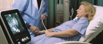
When it comes to diagnosing gallbladder problems, ultrasound is often the first imaging test that doctors turn to. This noninvasive procedure uses sound waves to create detailed images of the gallbladder and surrounding structures.
Once the ultrasound is completed, a radiologist will examine the images and provide a report that outlines the findings. Understanding the results of a gallbladder ultrasound can help patients and their doctors determine the best course of action for treatment.
One of the key things that the ultrasound report will indicate is the presence of gallstones. Gallstones are small, hardened deposits that can form in the gallbladder. They can cause symptoms such as pain, nausea, and vomiting. The ultrasound report will describe the size and location of any gallstones that are found.
In addition to looking for gallstones, the ultrasound can also reveal other conditions that may be affecting the gallbladder. The report may mention the presence of gallbladder inflammation, which is known as cholecystitis. It may also indicate the presence of gallbladder polyps, which are growths that can develop on the inner lining of the gallbladder.
Key Metrics Analyzed
When analyzing gallbladder ultrasound results, there are several key metrics that are typically examined. These metrics provide important information about the health and function of the gallbladder. Some of the key metrics that are commonly analyzed include:
- Gallbladder wall thickness: The thickness of the gallbladder wall is an important indicator of inflammation or infection. A thickened gallbladder wall may indicate conditions such as cholecystitis or gallbladder cancer.
- Gallbladder size: The size of the gallbladder can vary depending on factors such as age, sex, and body size. Abnormally large or small gallbladders may indicate underlying health conditions.
- Gallstones: Gallstones are solid deposits that can form in the gallbladder. The presence of gallstones can indicate gallbladder disease or other conditions.
- Biliary sludge: Biliary sludge refers to a mixture of cholesterol crystals, calcium salts, and other substances that can accumulate in the gallbladder. The presence of biliary sludge may indicate an increased risk of gallstones or other gallbladder problems.
- Gallbladder contractility: The ability of the gallbladder to contract and empty bile is an important measure of its overall function. Impaired gallbladder contractility may indicate gallbladder disease or dysfunction.
- Ductal dilation: Ductal dilation refers to the enlargement of the bile ducts. This can be a sign of gallbladder or bile duct obstruction.
By analyzing these key metrics, healthcare providers can gain valuable insights into the health and function of the gallbladder. This information can help guide diagnosis and treatment decisions for patients with gallbladder conditions.
Gallbladder Size and Shape
The size and shape of the gallbladder are important factors that can be assessed during a gallbladder ultrasound. The normal size of the gallbladder can vary, but it is typically around 7 to 10 centimeters in length and 3 to 5 centimeters in diameter. However, these measurements can vary depending on a person’s age, body size, and other factors.
During a gallbladder ultrasound, the shape of the gallbladder is also evaluated. A normal gallbladder typically has a pear-like shape, with a rounded bottom and a narrower neck. However, variations in shape can occur, and these can sometimes be indicative of certain conditions.
For example, if the gallbladder appears elongated or distended, this could be a sign of gallbladder obstruction or inflammation. On the other hand, if the gallbladder appears contracted or shrunken, this could indicate chronic gallbladder disease or a history of gallstones.
It is important to note that gallbladder size and shape alone cannot provide a definitive diagnosis. They are just one piece of the puzzle and must be evaluated in conjunction with other ultrasound findings, clinical symptoms, and laboratory tests to make an accurate diagnosis.
Overall, a gallbladder ultrasound can provide valuable information about the size and shape of the gallbladder, which can help healthcare providers in evaluating the overall health of the gallbladder and identifying any abnormalities that may be present.
Gallstones Detected
If your gallbladder ultrasound shows the presence of gallstones, it means that small, hard deposits have formed in your gallbladder. Gallstones are quite common and can vary in size and number. They are typically made up of cholesterol or bilirubin, a pigment produced by the liver.
Gallstones can be asymptomatic and may not require treatment. However, if they cause symptoms such as abdominal pain, bloating, nausea, or vomiting, your doctor may recommend treatment options. These may include medication to dissolve the stones, or surgical procedures such as laparoscopic cholecystectomy to remove the gallbladder.
It’s important to follow up with your doctor to discuss the implications of gallstones and determine the best course of action based on your specific situation. They will be able to provide you with personalized guidance and recommendations.
Bile Flow Assessment
During a gallbladder ultrasound, the technician may also assess the flow of bile. Bile is a digestive fluid produced by the liver and stored in the gallbladder. It helps in the digestion and absorption of fats.
The technician will look for any signs of obstruction or blockage in the bile ducts. This can be caused by gallstones, tumors, or other abnormalities. If the bile flow is blocked, it can lead to symptoms such as jaundice, abdominal pain, and digestive problems.
The ultrasound can help determine the cause and severity of the blockage. It can also help identify any potential complications, such as inflammation or infection of the bile ducts.
If a blockage is detected, further tests or procedures may be recommended to determine the appropriate treatment. This may include additional imaging tests, blood tests, or even surgery.
Overall, assessing the bile flow is an important part of a gallbladder ultrasound. It can help diagnose and manage various conditions related to the gallbladder and bile ducts.
Common Gallbladder Abnormalities
Gallbladder ultrasound results can reveal various abnormalities that may indicate potential health issues or conditions affecting the gallbladder. Some common abnormalities identified during a gallbladder ultrasound include:
Gallstones: Gallstones are one of the most common abnormalities detected during a gallbladder ultrasound. These are hard deposits that form in the gallbladder and can cause pain, inflammation, and other complications. Gallstones may appear as bright echoes on the ultrasound image.
Gallbladder Polyps: Polyps are abnormal growths that can develop on the inner lining of the gallbladder. While most polyps are benign, some can be cancerous or have the potential to become cancerous. Ultrasound can help detect the presence of gallbladder polyps and determine their size and characteristics.
Gallbladder Wall Thickening: Thickening of the gallbladder wall can indicate inflammation or infection, such as cholecystitis or gallbladder infection. This condition can cause abdominal pain, fever, and other symptoms. Ultrasound can measure the thickness of the gallbladder wall and help diagnose the underlying cause.
Gallbladder Sludge: Gallbladder sludge refers to a thickened mixture of bile and other substances that can accumulate in the gallbladder. Sludge may be a precursor to the formation of gallstones and can cause similar symptoms. Ultrasound can detect the presence of sludge and help guide further treatment.
Gallbladder Tumors: While rare, gallbladder tumors can be detected through ultrasound imaging. Tumors can be benign or malignant and may require further testing and treatment. Ultrasound can provide initial information about the size, location, and characteristics of the tumor.
Gallbladder Polypoid Lesions: Polypoid lesions are abnormal growths that resemble polyps but have different characteristics. These lesions can be benign or malignant and may require further evaluation. Ultrasound can help identify and evaluate gallbladder polypoid lesions.
Gallbladder Dysfunction: In some cases, gallbladder ultrasound may reveal abnormalities in the functioning of the gallbladder, such as poor contraction or impaired emptying. These dysfunctions can contribute to symptoms like pain and digestive issues. Ultrasound can help assess the gallbladder’s functionality and guide further treatment.
It is important to note that the interpretation of gallbladder ultrasound results should be done by a qualified medical professional. They will consider the individual’s medical history, symptoms, and other diagnostic tests to reach an accurate diagnosis and recommend appropriate treatment.
Cholecystitis
Cholecystitis is a condition characterized by inflammation of the gallbladder. It is often caused by the presence of gallstones blocking the bile ducts, leading to an accumulation of bile and subsequent irritation of the gallbladder walls.
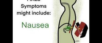
Common symptoms of cholecystitis include severe abdominal pain, fever, nausea, and vomiting. The pain is typically located in the upper right quadrant of the abdomen and may radiate to the shoulder or back.
During a gallbladder ultrasound, signs of cholecystitis may be observed. These include thickening of the gallbladder wall, fluid accumulation around the gallbladder, and the presence of gallstones. The ultrasound may also reveal an enlarged gallbladder and signs of infection, such as a buildup of pus.
Treatment for cholecystitis often involves surgical removal of the gallbladder, known as a cholecystectomy. This is typically performed laparoscopically, using small incisions and a camera-guided instrument. In cases where surgery is not an option, medications may be prescribed to manage symptoms and prevent complications.
If cholecystitis is left untreated, it can lead to serious complications, such as a ruptured gallbladder or the development of an abscess. Therefore, it is important to seek medical attention if you experience symptoms of cholecystitis or if your gallbladder ultrasound shows signs of inflammation.
| Symptoms of Cholecystitis |
| – Severe abdominal pain |
| – Fever |
| – Nausea |
| – Vomiting |
Gallstones
Gallstones are hardened deposits that form in the gallbladder, a small organ located beneath the liver. These stones can vary in size and shape and are typically made up of cholesterol or bilirubin, a substance produced during the breakdown of red blood cells.
Gallstones can develop when there is an imbalance in the chemicals present in the bile, a digestive fluid that helps break down fats. When the bile contains too much cholesterol or bilirubin, it can crystallize and form stones.
Most people with gallstones do not experience symptoms and may not be aware of their presence. However, if a gallstone becomes lodged in a bile duct, it can cause severe pain, known as a gallbladder attack. Symptoms of a gallbladder attack may include pain in the upper abdomen, nausea, vomiting, and fever.
Gallstones can be diagnosed through a gallbladder ultrasound, which uses sound waves to create images of the gallbladder and surrounding structures. The ultrasound can determine the size, number, and location of the gallstones.
Treatment for gallstones may be necessary if they are causing symptoms or complications. In some cases, medication can be prescribed to dissolve the stones. However, if the gallstones are causing severe pain or other complications, surgical removal of the gallbladder may be necessary.
Prevention of gallstones can be achieved by maintaining a healthy weight, eating a balanced diet high in fiber and low in cholesterol, and staying hydrated. Regular exercise and avoiding rapid weight loss can also help reduce the risk of developing gallstones.
Polyps and Tumors
During a gallbladder ultrasound, polyps and tumors may be detected. Polyps are small growths that can develop on the lining of the gallbladder. While most polyps are benign and do not cause any symptoms, some larger polyps have the potential to become cancerous.
If a polyp is detected during the ultrasound, the size, shape, and location of the polyp will be evaluated. Depending on the characteristics of the polyp, further testing or monitoring may be recommended. In some cases, a biopsy may be performed to determine if the polyp is cancerous.
Tumors in the gallbladder can also be detected during an ultrasound. Tumors can be either benign or malignant. Benign tumors are non-cancerous and generally do not pose a threat to health. Malignant tumors, on the other hand, are cancerous and can spread to other parts of the body.
If a tumor is detected, further testing will be needed to determine if it is benign or malignant. This may include additional imaging tests, such as a CT scan or MRI, or a biopsy to examine cells from the tumor. Treatment options for gallbladder tumors will depend on whether they are benign or malignant and may include surgery, chemotherapy, or radiation therapy.
It is important to note that not all gallbladder polyps or tumors require treatment. In many cases, small benign polyps may be monitored over time to ensure they do not grow or become cancerous. However, if there is concern about the potential for cancer or if symptoms are present, further evaluation and treatment may be necessary.
It is essential to consult with a healthcare professional to interpret gallbladder ultrasound results and determine the appropriate course of action.
Treatment Options for Detected Problems
Once a problem has been detected through a gallbladder ultrasound, there are several treatment options that may be recommended by your healthcare provider. The specific treatment will depend on the nature and severity of the problem.
If gallstones are detected, your doctor may recommend medications to help dissolve the stones or surgical removal of the gallbladder. Medications such as ursodeoxycholic acid may be prescribed to dissolve smaller gallstones over time. However, this treatment option is typically only effective for certain types of stones and may take several months to work.
If the gallstones are causing severe symptoms or complications, surgical removal of the gallbladder may be necessary. This procedure, known as a cholecystectomy, can be done laparoscopically or through open surgery. Laparoscopic cholecystectomy is less invasive and has a shorter recovery time compared to open surgery.
If the ultrasound reveals inflammation of the gallbladder (cholecystitis), treatment may involve pain medication, antibiotics to treat any infection, and a low-fat diet. In some cases, a cholecystectomy may be recommended to prevent future episodes of cholecystitis.
In rare cases, more serious conditions such as gallbladder cancer or bile duct obstruction may be detected through a gallbladder ultrasound. The treatment for these conditions will depend on the stage and severity of the disease and may involve surgery, radiation therapy, or chemotherapy.
It is important to discuss the treatment options with your healthcare provider to determine the best course of action based on your individual situation. They will consider factors such as the specific problem detected, your overall health, and any other underlying conditions you may have.
| Treatment Options | Description |
| Medication | Prescribed to dissolve gallstones or manage symptoms |
| Surgical Removal of Gallbladder | Recommended for severe gallstones or complications |
| Pain Medication and Antibiotics | Treatment for cholecystitis and infection |
| Low-Fat Diet | Recommended for cholecystitis and gallstones |
| Surgery, Radiation Therapy, or Chemotherapy | Treatment options for more serious conditions |
Surgery for Gallstones
If gallstones are causing severe symptoms or complications, surgery may be recommended to remove the gallbladder. This procedure is known as a cholecystectomy. There are two main types of cholecystectomy:
- Laparoscopic cholecystectomy: This is the most common type of surgery for gallstones. It involves making several small incisions in the abdomen and inserting a tiny camera and surgical instruments to remove the gallbladder. Laparoscopic cholecystectomy is minimally invasive and typically has a shorter recovery time compared to open surgery.
- Open cholecystectomy: In some cases, open surgery may be necessary, especially if there are complications or if the gallbladder is severely inflamed or infected. This procedure involves making a larger incision in the abdomen to remove the gallbladder. Open cholecystectomy may require a longer hospital stay and recovery time.
Both types of cholecystectomy are generally safe and effective in treating gallstones. The surgeon will evaluate the individual’s condition and determine the most appropriate approach. After the gallbladder is removed, bile will flow directly from the liver into the small intestine.
It is important to discuss the risks, benefits, and potential complications of surgery with a healthcare professional. Some possible risks include infection, bleeding, injury to surrounding organs, and bile duct injury. In rare cases, bile leakage or the formation of a bile duct stone may occur after surgery.
After surgery, most individuals can resume their normal activities within a few days to a week. They may need to follow a special diet for a short period of time to aid in digestion. In the absence of a gallbladder, the body can still function normally, as the liver will continue to produce bile to aid in the digestion of fats.
Overall, surgery for gallstones is a common and effective treatment option. It can provide relief from symptoms and reduce the risk of complications associated with gallstones.
Medications for Inflammation
There are several medications that can be used to treat inflammation, including:
- Nonsteroidal anti-inflammatory drugs (NSAIDs): These drugs help reduce inflammation and relieve pain. Examples include ibuprofen, naproxen, and aspirin. NSAIDs can have side effects and should be used with caution, especially in people with certain medical conditions.
- Corticosteroids: These medications are powerful anti-inflammatory drugs that can be taken orally, injected, or applied topically. They work by suppressing the immune system and reducing inflammation. Corticosteroids can have significant side effects and should be used under the guidance of a healthcare professional.
- Disease-modifying antirheumatic drugs (DMARDs): These drugs are used to treat chronic inflammatory conditions, such as rheumatoid arthritis. DMARDs work by targeting the underlying immune system dysfunction that causes inflammation. Examples include methotrexate, sulfasalazine, and hydroxychloroquine.
- Biologic agents: These are a newer class of medications that are used to treat certain inflammatory conditions, such as rheumatoid arthritis and psoriasis. Biologic agents work by targeting specific molecules in the immune system that are involved in the inflammatory response. Examples include adalimumab, etanercept, and infliximab.
- Immunosuppressants: These medications are used to suppress the immune system and reduce inflammation. They are often used in the treatment of autoimmune diseases, such as lupus and Crohn’s disease. Examples include azathioprine, cyclosporine, and mycophenolate mofetil.
It is important to note that these medications should only be used under the guidance of a healthcare professional. They can have side effects and may interact with other medications. Your doctor will determine the most appropriate medication for your specific condition and monitor your response to treatment.
Follow-up Scans for Monitoring
After receiving the initial gallbladder ultrasound results, your healthcare provider may recommend follow-up scans for monitoring purposes. These additional scans are typically performed to track any changes in the gallbladder over time and to assess the effectiveness of any treatment or interventions that have been implemented.
During a follow-up scan, the sonographer will use the same ultrasound technology to obtain images of the gallbladder. The images will be compared to the previous ultrasound results to look for any new abnormalities or changes in the gallbladder’s structure or function.
The frequency of follow-up scans will depend on various factors, including the specific condition being monitored and the patient’s individual circumstances. Some individuals may only require periodic monitoring, while others may need more frequent scans to closely track their condition.
Follow-up scans can provide valuable information about the progression or regression of gallbladder conditions, such as gallstones, inflammation, or tumors. They can help determine if the treatment plan is working effectively or if any adjustments need to be made.
If any new abnormalities or changes are identified during a follow-up scan, further diagnostic tests or interventions may be recommended. These may include additional imaging tests, such as a CT scan or an MRI, or more invasive procedures, such as a biopsy or surgery.
| Benefits of Follow-up Scans | Risks and Considerations |
|
|
It is important to follow your healthcare provider’s recommendations for follow-up scans to ensure the ongoing monitoring and management of your gallbladder condition. These scans can provide valuable insight into the progression of the condition and help guide treatment decisions.
FAQ:
What are gallbladder ultrasound results?
Gallbladder ultrasound results refer to the findings obtained from an ultrasound examination of the gallbladder. This imaging technique uses sound waves to create images of the gallbladder and surrounding structures, allowing doctors to evaluate the organ’s size, shape, and overall condition.
What can gallbladder ultrasound results reveal?
Gallbladder ultrasound results can reveal various conditions and abnormalities, such as gallstones, inflammation of the gallbladder (cholecystitis), gallbladder polyps, tumors, or other structural abnormalities. The results can also show if the gallbladder is functioning properly or if there are any signs of obstruction or blockage in the bile ducts.
Are gallstones always detected in gallbladder ultrasound results?
No, gallstones are not always detected in gallbladder ultrasound results. Although ultrasound is a commonly used imaging technique for detecting gallstones, small stones or those located deep within the gallbladder may not be visible on the ultrasound images. In such cases, other imaging tests, such as a CT scan or MRI, may be recommended to confirm the presence of gallstones.
What does it mean if gallbladder ultrasound results show inflammation?
If gallbladder ultrasound results show inflammation, it may indicate a condition called cholecystitis. Cholecystitis is often caused by gallstones blocking the bile ducts or by an infection in the gallbladder. Inflammation of the gallbladder can cause symptoms such as abdominal pain, fever, and nausea. Treatment may involve medication to relieve symptoms, antibiotics to treat infection, or surgery to remove the gallbladder.
Can gallbladder ultrasound results detect cancer?
Yes, gallbladder ultrasound results can sometimes detect cancerous tumors in the gallbladder. The ultrasound images may show abnormal growths or masses in the gallbladder that could be indicative of cancer. However, a definitive diagnosis of gallbladder cancer usually requires further testing, such as a biopsy, to analyze the cells for cancerous changes. It’s important to consult with a healthcare professional for an accurate diagnosis and appropriate treatment if cancer is suspected.
What is a gallbladder ultrasound?
A gallbladder ultrasound is a non-invasive imaging test that uses sound waves to create pictures of the gallbladder. It is used to evaluate the gallbladder for any abnormalities or diseases.
What are some common reasons for getting a gallbladder ultrasound?
Some common reasons for getting a gallbladder ultrasound include abdominal pain, jaundice, abnormal liver function tests, suspected gallstones, or to monitor the progression of a known gallbladder disease.
What can the results of a gallbladder ultrasound indicate?
The results of a gallbladder ultrasound can indicate various conditions such as gallstones, gallbladder inflammation (cholecystitis), gallbladder polyps, gallbladder sludge, or gallbladder cancer.
What are the different types of gallbladder abnormalities that can be detected through ultrasound?
Some of the different types of gallbladder abnormalities that can be detected through ultrasound include gallstones, thickening of the gallbladder wall, dilated bile ducts, and presence of fluid around the gallbladder.
What do the different ultrasound findings mean?
The ultrasound findings can help diagnose specific conditions. For example, the presence of gallstones can indicate cholelithiasis, while thickening of the gallbladder wall can suggest cholecystitis. The interpretation of the findings depends on the patient’s symptoms and medical history.