
Ultrasound Evaluations: Can MALS be Missed?
MALS (Median Arcuate Ligament Syndrome) is a rare but potentially debilitating condition that affects the blood flow to the digestive organs. It occurs when the median arcuate ligament, which is located just below the diaphragm, compresses the celiac artery.
Due to its rarity and non-specific symptoms, MALS can often be missed or misdiagnosed. Patients may present with abdominal pain, weight loss, and nausea, which are symptoms that can be attributed to a variety of other conditions. For those facing such symptoms and suspecting MALS, understanding the available Mals Syndrome Treatment options is paramount. However, an accurate diagnosis is crucial as untreated MALS can lead to serious complications.
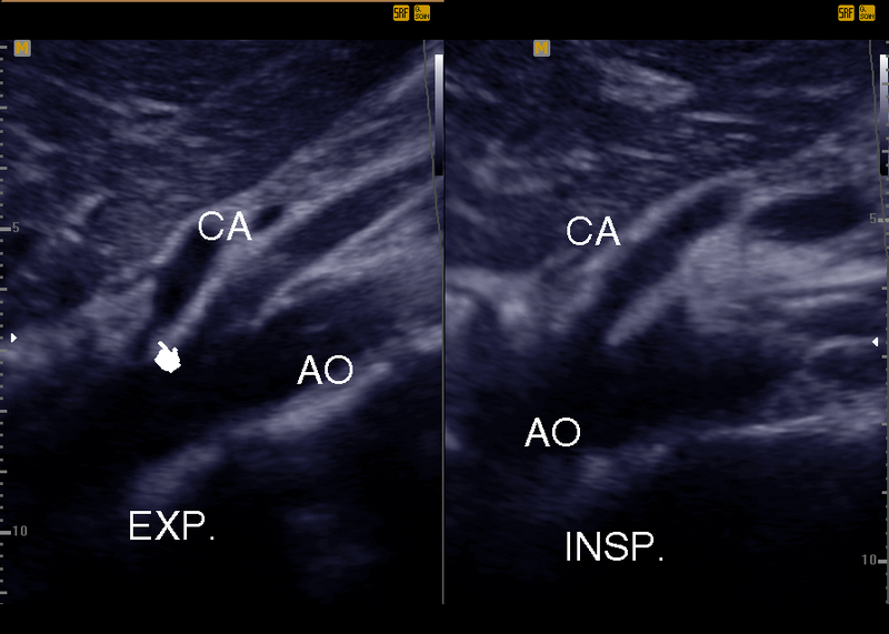
One of the most reliable and non-invasive methods to diagnose MALS is through ultrasound evaluations. Ultrasound uses sound waves to create images of the body’s internal structures, allowing physicians to visualize any abnormalities or blockages in the celiac artery. This diagnostic technique is safe, cost-effective, and readily available in most medical facilities.
In recent years, the importance of ultrasound evaluations in diagnosing Median Arcuate Ligament Syndrome has become increasingly recognized. By detecting the compression of the celiac artery, ultrasound can provide an accurate diagnosis and guide appropriate treatment options. Furthermore, ultrasound evaluations can also be used to assess the effectiveness of the chosen treatment and monitor the patient’s progress.
Median Arcuate Ligament Syndrome is a condition that can easily be missed due to its rarity and non-specific symptoms. However, with the advancements in ultrasound technology, physicians now have a reliable and accessible tool to diagnose and manage MALS effectively. Therefore, ultrasound evaluations play a critical role in the early detection and treatment of this condition.