 The Importance of Ultrasound in the Diagnosis of Celiac Disease: A Comprehensive Guide to its Role in Identifying and Managing the Condition
Celiac disease is a chronic autoimmune disorder that affects the small intestine and is triggered by the consumption of gluten, a protein found in wheat, barley, and rye. It is estimated to affect 1 in 100 people worldwide. The diagnosis of celiac disease typically involves a combination of clinical evaluation, serological tests, and duodenal biopsy. However, ultrasound imaging has emerged as a valuable tool in the diagnosis and monitoring of celiac disease.
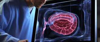
Ultrasound, also known as sonography, is a non-invasive imaging technique that uses high-frequency sound waves to produce images of the body’s internal structures. In the case of celiac disease, ultrasound can be used to assess the small intestine for characteristic findings such as thickening of the intestinal wall, dilated loops of bowel, and fluid accumulation.
One of the advantages of ultrasound in diagnosing celiac disease is its ability to visualize the small intestine in real time, allowing for immediate assessment of the intestinal wall and surrounding structures. This can help differentiate between celiac disease and other conditions that may present with similar symptoms, such as inflammatory bowel disease or small bowel obstruction.
In addition to diagnosing celiac disease, ultrasound can also be used to monitor the progression of the disease and assess response to treatment. Serial ultrasound examinations can detect changes in the intestinal wall thickness and other signs of disease activity, providing valuable information for guiding treatment decisions.
Ultrasound imaging plays a crucial role in the diagnosis and management of celiac disease. Its ability to visualize the small intestine and assess for characteristic findings makes it a valuable tool in the diagnostic workup of patients with suspected celiac disease. Furthermore, serial ultrasound examinations can provide important information for monitoring disease progression and guiding treatment decisions. As our understanding of celiac disease continues to evolve, ultrasound will likely play an even greater role in its diagnosis and management.
Potential Markers of Celiac Disease
There are several potential markers that can help in the diagnosis of celiac disease. These markers can indicate the presence of the disease and help healthcare professionals make an accurate diagnosis. Some of the potential markers include:
- Anti-tissue transglutaminase antibodies (anti-tTG): These antibodies are commonly found in individuals with celiac disease. Testing for anti-tTG antibodies can help identify the presence of the disease.
- Anti-endomysial antibodies (EMA): Similar to anti-tTG antibodies, EMA antibodies are often elevated in individuals with celiac disease. Testing for EMA antibodies can be another useful marker in the diagnosis process.
- Anti-deamidated gliadin peptides (anti-DGP): These antibodies target a protein present in gluten and can be a marker of celiac disease. Testing for anti-DGP antibodies can provide additional evidence of the disease.
- HLA-DQ2 and HLA-DQ8 genotypes: These genetic markers are strongly associated with celiac disease. Testing for the presence of HLA-DQ2 and HLA-DQ8 genotypes can help confirm the diagnosis of the disease.
- Elevated levels of certain biomarkers: Individuals with celiac disease may have elevated levels of certain biomarkers, such as IgA, IgG, or fecal calprotectin. Testing for these biomarkers can provide further evidence of the disease.
It’s important to note that the presence of these potential markers does not definitively diagnose celiac disease. A combination of clinical evaluation, serological testing, and, in some cases, intestinal biopsy is necessary for an accurate diagnosis. However, these markers can serve as valuable tools in the diagnostic process and help healthcare professionals make informed decisions about further testing and treatment options.
Bowel Wall Thickening
Bowel wall thickening is a common finding in patients with celiac disease. It is usually seen in the small intestine, particularly the duodenum and jejunum. Ultrasound can help in assessing the degree of bowel wall thickening and can provide valuable information for diagnosing celiac disease.
Thickening of the bowel wall is caused by the infiltration of inflammatory cells and the deposition of collagen. This can lead to a loss of normal bowel architecture and can result in malabsorption of nutrients.
Ultrasound can detect bowel wall thickening by visualizing the layers of the bowel wall. The normal bowel wall consists of several layers, including the mucosa, submucosa, muscularis propria, and serosa. In patients with celiac disease, the bowel wall may appear thickened and have a “target” or “double halo” appearance.
In addition to assessing the degree of bowel wall thickening, ultrasound can also help in evaluating other features of celiac disease, such as the presence of lymphadenopathy and the appearance of the mesentery. Lymphadenopathy is often seen in celiac disease and can be detected as enlarged lymph nodes adjacent to the bowel wall. The mesentery may also appear thickened and hypervascular.
Overall, ultrasound is a valuable tool in diagnosing celiac disease and assessing the degree of bowel wall thickening. It can provide important information for guiding further diagnostic workup and treatment planning.
Reduced Peristalsis
Reduced peristalsis refers to the decreased movement of the muscles in the gastrointestinal tract, specifically the small intestine. This condition can be observed through ultrasound imaging and is often associated with celiac disease.
Ultrasound can be used to evaluate the motility of the small intestine and determine the presence of reduced peristalsis. This is done by observing the movement of the intestinal walls and the passage of food and fluid through the digestive system.
In patients with celiac disease, reduced peristalsis may be present due to the inflammation and damage to the lining of the small intestine. This can lead to a variety of symptoms, including abdominal pain, bloating, and diarrhea.
By detecting reduced peristalsis through ultrasound, healthcare providers can better diagnose celiac disease and monitor its progression. This information can be used to develop personalized treatment plans and improve patient outcomes.
Ultrasound plays a crucial role in diagnosing celiac disease by identifying the presence of reduced peristalsis. This non-invasive imaging technique provides valuable information about the motility of the small intestine and helps healthcare providers make informed decisions regarding patient care.
Enlarged Mesenteric Lymph Nodes
Enlarged mesenteric lymph nodes are a common finding in patients with celiac disease. These lymph nodes are located in the mesentery, which is the tissue that connects the intestines to the abdominal wall. They play an important role in the immune response and can become enlarged in response to inflammation or infection.
In patients with celiac disease, the immune system reacts abnormally to gluten, a protein found in wheat, barley, and rye. This abnormal immune response leads to inflammation in the small intestine, which can cause a variety of symptoms and damage to the intestinal lining.
When the immune system is activated in response to gluten, the mesenteric lymph nodes can become enlarged. This enlargement can be detected using ultrasound imaging. Ultrasound is a non-invasive imaging technique that uses sound waves to create detailed images of the body’s internal structures.
During an ultrasound examination, the ultrasound technician will place a small probe on the abdomen. The probe emits sound waves, which bounce off the internal organs and tissues and create echoes. These echoes are then converted into images that can be viewed on a monitor.
Enlarged mesenteric lymph nodes may appear as round or oval-shaped structures on the ultrasound images. They can vary in size and may be accompanied by other signs of inflammation, such as thickening of the intestinal wall.
Ultrasound imaging can help diagnose celiac disease by providing visual evidence of inflammation and enlarged mesenteric lymph nodes. It can also help monitor the response to treatment and detect any complications, such as abscesses or perforations, that may occur as a result of the disease.
However, it is important to note that ultrasound imaging alone is not sufficient to diagnose celiac disease. A definitive diagnosis requires a combination of clinical evaluation, blood tests, and intestinal biopsy.
Enlarged mesenteric lymph nodes are a common finding in patients with celiac disease and can be detected using ultrasound imaging. This non-invasive technique provides valuable information about the extent of inflammation and can aid in the diagnosis and management of the disease.
Advantages of Ultrasound
- Non-invasive: Ultrasound is a non-invasive imaging technique, meaning it does not require any incisions or the use of radiation. This makes it a safer option compared to other imaging methods such as CT scans or x-rays.
- Painless: Ultrasound procedures are painless and generally well-tolerated by patients. There is no discomfort associated with the use of ultrasound.
- Real-time imaging: Ultrasound provides real-time images, allowing the healthcare provider to immediately visualize the area of interest. This can help in quickly diagnosing celiac disease and monitoring the disease progression.
- No radiation exposure: Unlike other imaging techniques, ultrasound does not involve the use of radiation. This makes it a safer option, especially for patients who may need repeated imaging studies.
- Cost-effective: Ultrasound is a relatively cost-effective imaging modality compared to other methods such as MRI or CT scans. It can help reduce healthcare costs while still providing valuable diagnostic information.
- Portable: Ultrasound machines are portable and can be easily transported to different locations. This makes it convenient for patients who may not have easy access to specialized imaging centers.
- No known side effects: Ultrasound is generally considered safe, and there are no known side effects associated with its use. It can be safely used in patients of all ages, including children and pregnant women.
Non-Invasive
Ultrasound is a non-invasive imaging technique that uses sound waves to create images of the inside of the body. It is a safe and painless procedure that does not require the use of radiation or contrast agents, making it an ideal tool for diagnosing celiac disease.
One of the main advantages of ultrasound is its ability to assess the structure and function of the small intestine. By using high-frequency sound waves, ultrasound can provide detailed images of the small intestine, including the presence of any abnormalities or damage.
In the case of celiac disease, ultrasound can help detect certain signs that are indicative of the condition. These signs include thickening of the intestinal wall, dilated loops of the small intestine, and the presence of fluid-filled sacs called pseudocysts.
Furthermore, ultrasound can also be used to assess the presence of complications related to celiac disease, such as intestinal strictures or obstructions. This can be particularly helpful in monitoring the progression of the disease and guiding treatment decisions.
In addition to its diagnostic capabilities, ultrasound can also be used to guide certain procedures related to celiac disease, such as biopsies. By using ultrasound to visualize the area of interest, healthcare providers can ensure accurate and targeted biopsies, minimizing the risk of complications.
Overall, ultrasound is a valuable non-invasive tool in the diagnosis and management of celiac disease. Its ability to provide detailed images of the small intestine and detect signs of the condition makes it an important adjunct to other diagnostic methods, such as blood tests and endoscopy.
Wide Availability
One of the key advantages of using ultrasound in diagnosing celiac disease is its wide availability. Unlike other diagnostic tools such as endoscopy or biopsy, ultrasound machines are commonly found in medical facilities of all sizes, from small clinics to large hospitals.
This accessibility makes ultrasound a convenient option for both patients and healthcare providers. Patients can have the procedure done at a location that is convenient for them, without the need for referrals or long waiting times. Healthcare providers can easily incorporate ultrasound into their diagnostic practices, reducing the need for more invasive procedures and potentially lowering costs.
Furthermore, the wide availability of ultrasound allows for timely diagnosis and treatment of celiac disease. With quick access to ultrasound machines, healthcare providers can promptly evaluate patients who present with symptoms suggestive of celiac disease. Early diagnosis is crucial in managing the condition and preventing complications, and ultrasound can play a valuable role in facilitating this process.
No Radiation Exposure
One of the major advantages of using ultrasound in diagnosing celiac disease is that it does not involve any radiation exposure. Unlike other imaging techniques such as X-rays and CT scans, ultrasound uses sound waves to create images of the internal organs.
This is particularly important for patients with celiac disease, as they may need to undergo multiple imaging tests over time to monitor the progression of the disease or to check for complications. By using ultrasound, doctors can minimize the cumulative radiation dose that patients receive, reducing the potential risk of long-term radiation-related side effects.
In addition, ultrasound is a non-invasive procedure, meaning that it does not require any incisions or injections. This makes it a safer and more comfortable option for patients, especially children and individuals who may be sensitive to radiation or have a fear of medical procedures.
Furthermore, ultrasound is a relatively quick and convenient imaging technique. It can be performed in a doctor’s office or clinic setting, and the results are typically available immediately. This allows for faster diagnosis and treatment planning, which is crucial in managing celiac disease and its associated complications.
Overall, the absence of radiation exposure in ultrasound imaging makes it a valuable tool in diagnosing celiac disease, providing a safe and effective alternative to other imaging modalities.
Limitations of Ultrasound
While ultrasound is a valuable tool in diagnosing celiac disease, it does have certain limitations that should be taken into consideration.
Firstly, ultrasound is highly operator-dependent. The quality and accuracy of the ultrasound images can vary depending on the skill and experience of the sonographer. It is important to have a skilled sonographer who is familiar with the specific imaging techniques used for celiac disease diagnosis.
Additionally, ultrasound may not be able to detect all cases of celiac disease. It is most effective in identifying severe cases with significant changes in the intestinal walls. Mild or early-stage cases may not show noticeable abnormalities on ultrasound images.
Furthermore, ultrasound cannot provide a definitive diagnosis of celiac disease. It can only suggest the presence of certain characteristic findings that are associated with the disease. A definitive diagnosis can only be made through a combination of clinical symptoms, serologic tests, and histopathologic examination of a biopsy sample.
Finally, ultrasound may not be suitable for certain patient populations, such as those with obesity or excessive bowel gas. These factors can make it more difficult to obtain clear and accurate ultrasound images of the intestines.
Despite these limitations, ultrasound remains a valuable tool in the diagnostic process of celiac disease. It can provide additional information to support the clinical suspicion of the disease and help guide further diagnostic testing and treatment decisions.
Operator Dependent
It is important to note that the accuracy and reliability of ultrasound in diagnosing celiac disease can be highly dependent on the skill and experience of the operator performing the exam. Ultrasound is an operator-dependent imaging modality, meaning that the quality of the images and the interpretation of the findings can vary depending on the expertise of the person conducting the examination.
Operators who are experienced in performing ultrasound examinations for celiac disease are more likely to accurately identify and characterize the specific ultrasound features associated with the disease. They are also more likely to be familiar with the potential pitfalls and limitations of ultrasound in this context.
Conversely, inexperienced operators may struggle to obtain high-quality images or may misinterpret the findings, leading to inaccurate or inconclusive results. Therefore, it is crucial to have well-trained and experienced sonographers or radiologists who specialize in gastrointestinal ultrasound performing these exams.
| Benefits of Operator Expertise | Limitations of Inexperienced Operators |
| Accurate identification of ultrasound features associated with celiac disease | Difficulty obtaining high-quality images |
| Familiarity with potential pitfalls and limitations of ultrasound in this context | Potential misinterpretation of findings |
| Ability to provide more accurate and reliable diagnoses | Possible generation of inconclusive or inaccurate results |
Overall, the operator’s expertise and experience play a crucial role in the effectiveness of ultrasound in diagnosing celiac disease. Collaborating with skilled operators can improve the accuracy and reliability of the ultrasound examination, leading to more confident diagnoses and better patient outcomes.
Lower Sensitivity for Mild Cases
While ultrasound can be an effective tool for diagnosing celiac disease, it does have limitations, particularly when it comes to detecting mild cases. In these instances, ultrasound may not be as sensitive in identifying the characteristic changes in the small intestine that are indicative of celiac disease.
One reason for this lower sensitivity is that mild cases of celiac disease may not present with significant structural abnormalities in the small intestine. Ultrasound relies on visualizing these abnormalities, such as blunting of the villi or a thickened bowel wall, to make a definitive diagnosis. Therefore, if these changes are not present or are minimal, ultrasound may not be able to accurately detect celiac disease.
Another factor that can affect the sensitivity of ultrasound in diagnosing celiac disease is the skill and experience of the sonographer. Ultrasound is a highly operator-dependent imaging technique, and the accuracy of the results can vary depending on the expertise of the person performing the procedure. A less experienced sonographer may have difficulty identifying subtle changes in the small intestine that could indicate celiac disease.
Despite these limitations, ultrasound can still play a valuable role in the diagnosis of celiac disease, particularly in cases where there are more pronounced structural abnormalities. It can also be useful in monitoring the response to treatment and assessing the progression of the disease over time.
| Advantages of Ultrasound in Diagnosing Celiac Disease | Limitations of Ultrasound in Diagnosing Celiac Disease |
| Non-invasive and painless procedure | Lower sensitivity for mild cases |
| No exposure to ionizing radiation | Operator-dependent nature of the procedure |
| Can provide real-time imaging | Relies on visualizing structural abnormalities |
Cannot Replace Biopsy
While ultrasound can be a useful tool in diagnosing celiac disease, it cannot replace the need for a biopsy. The gold standard for diagnosing celiac disease is still the histological examination of a small intestine biopsy sample. This involves the analysis of tissue samples taken from the small intestine to look for specific changes that are characteristic of celiac disease.
Ultrasound imaging can provide valuable information about the structure and appearance of the small intestine, but it cannot definitively diagnose celiac disease. Ultrasound can show signs of inflammation or other abnormalities that may be suggestive of celiac disease, but a biopsy is necessary to confirm the diagnosis.
The biopsy allows for a more detailed examination of the intestinal lining, providing a clearer picture of the extent of damage and the presence of any other related conditions. It can also help differentiate celiac disease from other conditions that may have similar symptoms.
It is worth noting that ultrasound can be a helpful tool in guiding the biopsy procedure. It can help the healthcare provider accurately locate the area of the small intestine that needs to be sampled, increasing the chances of obtaining an accurate diagnosis.
While ultrasound can provide valuable information and assist in the diagnosis of celiac disease, it cannot replace the need for a biopsy. The biopsy remains the gold standard for confirming the presence of celiac disease and assessing the extent of intestinal damage.
Ultrasound Protocol in Celiac Disease
Ultrasound imaging is a valuable tool in diagnosing celiac disease. It can provide important information about the small intestine, which is the primary site of damage in this condition. A standardized ultrasound protocol can help ensure accurate and consistent results in the diagnosis of celiac disease.
When performing an ultrasound for celiac disease, the following protocol is typically followed:
- Preparation: The patient should be fasting for at least six hours before the ultrasound to ensure optimal imaging of the small intestine.
- Positioning: The patient is positioned in a supine or left lateral decubitus position to allow for better visualization of the small intestine.
- Transducer: A high-frequency transducer, typically in the range of 5-10 MHz, is used to obtain detailed images of the small intestine.
- Scanning Technique: The transducer is placed on the abdomen and moved in a systematic manner to scan the entire small intestine. Both longitudinal and transverse views should be obtained to evaluate the entire length of the intestine.
- Image Interpretation: The sonographer or radiologist looks for specific findings that may indicate celiac disease, such as thickening of the intestinal wall, loss of normal folds, dilated loops of bowel, and fluid-filled segments.
- Reporting: The findings of the ultrasound are documented in a report, which is then shared with the referring physician for further evaluation and management.
It is important to note that while ultrasound can provide valuable information in the diagnosis of celiac disease, it is not the sole diagnostic tool. A combination of clinical history, serology, and histopathology is typically used to confirm the diagnosis. Ultrasound serves as a non-invasive imaging modality that can aid in the evaluation of celiac disease and guide further diagnostic and treatment decisions.
A standardized ultrasound protocol is essential in the diagnosis of celiac disease. Following a consistent approach ensures accurate and reliable results, helping clinicians make informed decisions about patient management.
Scanning Technique
Ultrasound scanning is a non-invasive imaging technique that uses high-frequency sound waves to produce detailed images of the internal organs and tissues. In the case of diagnosing celiac disease, ultrasound can be used to examine the small intestine for any abnormalities or signs of inflammation.
The scanning technique for celiac disease involves the use of a transducer, which is a handheld device that emits and receives sound waves. The transducer is placed on the abdomen and moved around to capture images of the small intestine.
During the scanning procedure, the patient may be asked to change positions or hold their breath to obtain clearer images. Gel is applied to the skin to ensure good contact between the transducer and the body, allowing the sound waves to travel effectively.
The images obtained during the ultrasound scan are displayed on a monitor in real-time, allowing the healthcare provider to assess the condition of the small intestine. The presence of abnormalities, such as thickening of the intestinal walls or dilated blood vessels, may indicate celiac disease.
It is important to note that ultrasound scanning is not the primary method for diagnosing celiac disease. It is typically used as a supplementary tool alongside other diagnostic tests, such as blood tests and endoscopy. However, ultrasound can provide valuable information about the state of the small intestine and assist in the diagnosis and management of celiac disease.
The scanning technique for celiac disease involves the use of ultrasound to examine the small intestine for any abnormalities or signs of inflammation. It is a non-invasive and safe procedure that can provide valuable information to aid in the diagnosis and management of celiac disease.
Measurement Parameters
Ultrasound can provide valuable information for diagnosing celiac disease by measuring various parameters. These parameters include:
Bowel Wall Thickness: The thickness of the bowel wall can be measured using ultrasound. In patients with celiac disease, the bowel wall is typically thickened due to inflammation. Measuring the bowel wall thickness can help confirm the presence of celiac disease.
Bowel Wall Layering: Ultrasound can also assess the layering of the bowel wall. In celiac disease, the layering pattern may be disrupted due to inflammation. This disruption can be visualized using ultrasound and can provide additional evidence for the diagnosis of celiac disease.
Lymph Node Size: Enlarged lymph nodes can be a sign of inflammation, which is commonly seen in celiac disease. Ultrasound can measure the size of lymph nodes in the abdomen and provide important information about the extent of inflammation in celiac disease.
Blood Flow: Doppler ultrasound can assess the blood flow in the bowel wall. In celiac disease, increased blood flow may be seen due to inflammation. This increased blood flow can be detected using Doppler ultrasound and can be a useful diagnostic tool for celiac disease.
Intestinal Motility: Ultrasound can also evaluate the motility of the intestines. In patients with celiac disease, the motility of the intestines may be affected due to inflammation and damage to the intestinal lining. Ultrasound can assess the movement of the intestines and provide information about the functionality of the gastrointestinal tract.
Other Findings: Ultrasound can detect other findings that may be associated with celiac disease, such as fluid accumulation in the abdomen (ascites) or abnormal masses. These findings can provide additional clues for the diagnosis of celiac disease.
Overall, ultrasound has proven to be a valuable tool for diagnosing celiac disease by measuring various parameters related to bowel wall thickness, layering, lymph node size, blood flow, intestinal motility, and other associated findings.
Image Evaluation
When using ultrasound to diagnose celiac disease, several key imaging criteria are evaluated. These criteria include:
- Bowel wall thickness: The thickness of the small bowel wall is measured and compared to normal values. Increased thickness can indicate inflammation and damage associated with celiac disease.
- Bowel wall echogenicity: The echogenicity, or brightness, of the bowel wall is assessed. Increased echogenicity can suggest the presence of fibrosis or scarring.
- Bowel wall stratification: The layers of the bowel wall are evaluated for any abnormalities. Disruption or irregularity in the layers can indicate inflammation or damage.
- Mesenteric lymphadenopathy: The presence of enlarged lymph nodes in the mesentery, the tissue that connects the small bowel to the abdominal wall, is assessed. Enlarged lymph nodes can be a sign of inflammation or infection.
- Peri-intestinal findings: The presence of fluid collections, abscesses, or other abnormalities around the small bowel is evaluated. These findings can indicate complications of celiac disease, such as perforation or abscess formation.
By carefully evaluating these imaging criteria, ultrasound can provide valuable information for the diagnosis and management of celiac disease.
Future Applications
As technology continues to advance, the role of ultrasound in diagnosing celiac disease is likely to expand. One potential future application is the use of ultrasound to monitor the effectiveness of treatment in celiac patients. Currently, the gold standard for assessing treatment response is repeat endoscopy with biopsy, which is invasive and carries risks. Ultrasound could provide a non-invasive alternative, allowing for regular monitoring of intestinal healing and response to treatment.
Another potential application is the use of ultrasound in screening for celiac disease. Currently, diagnosis often requires a combination of blood tests and endoscopy with biopsy. Ultrasound could potentially be used as a first-line screening tool, helping to identify individuals who may require further testing. This could lead to earlier diagnosis and treatment, improving outcomes for patients.
Additionally, ultrasound may have a role in assessing complications of celiac disease. For example, individuals with celiac disease are at increased risk for developing small bowel strictures, which can lead to obstruction. Ultrasound could be used to detect and monitor these strictures, allowing for early intervention and management.
The future applications of ultrasound in diagnosing celiac disease are promising. From monitoring treatment response to screening for the disease and assessing complications, ultrasound has the potential to improve patient care and outcomes. Continued research and technological advancements will be crucial in realizing these possibilities.
Screening and Monitoring
Ultrasound is a valuable tool for screening individuals at risk for celiac disease. It can help identify characteristic findings such as increased bowel wall thickness, reduced bowel motility, and enlarged mesenteric lymph nodes. These findings can suggest the presence of celiac disease and prompt further diagnostic testing.
Once a diagnosis of celiac disease has been confirmed, ultrasound can also play a role in monitoring the disease. Regular ultrasound examinations can help assess the response to treatment and provide information on disease progression or complications.
In addition to ultrasound, other diagnostic tools such as blood tests and endoscopy are commonly used for screening and monitoring celiac disease. However, ultrasound offers several advantages. It is non-invasive, widely available, and relatively low-cost compared to other imaging modalities. It also does not involve exposure to ionizing radiation, making it a safer option, particularly for children and pregnant women.
While ultrasound can provide valuable information, it is important to note that it is not a definitive diagnostic tool for celiac disease. A biopsy of the small intestine remains the gold standard for diagnosis. However, ultrasound can be a useful adjunct to help identify individuals at risk for celiac disease and monitor their condition over time.
Overall, ultrasound has a significant role in both screening and monitoring celiac disease. Its non-invasive nature, accessibility, and safety make it a valuable tool for healthcare professionals in the management of this chronic autoimmune disorder.
Paediatric Celiac Disease
Celiac disease, also known as gluten-sensitive enteropathy, is a chronic autoimmune disorder that affects the small intestine. It is triggered by the ingestion of gluten, a protein found in wheat, barley, and rye. Paediatric celiac disease refers to the condition when it is diagnosed in children.
Children with celiac disease often experience symptoms such as abdominal pain, diarrhea, bloating, and weight loss. However, some children may not exhibit any symptoms, making diagnosis challenging. It is important to identify celiac disease in children as early as possible to prevent complications and promote proper growth and development.
Ultrasound is a non-invasive imaging technique that can be used to diagnose and monitor paediatric celiac disease. It can help identify changes in the small intestine, such as thickening of the intestinal wall and dilation of the bowel loops. These findings can indicate inflammation and damage to the intestine, which are characteristic of celiac disease.
In addition to ultrasound, other diagnostic tools such as blood tests and endoscopy with biopsy may be used to confirm the diagnosis of paediatric celiac disease. Blood tests can detect specific antibodies that are present in celiac disease, while endoscopy with biopsy involves taking small samples of the intestinal tissue for analysis.
Ultrasound can also be used to monitor the response to treatment in children with celiac disease. Repeat ultrasound examinations can help assess the healing of the intestinal mucosa and the resolution of inflammation. This can guide the management of the disease and help determine the effectiveness of a gluten-free diet.
Ultrasound plays a valuable role in the diagnosis and monitoring of paediatric celiac disease. It is a non-invasive and safe imaging modality that can provide important information about the condition of the small intestine in children. Early detection and management of celiac disease are crucial to ensure optimal health and well-being in paediatric patients.
Point-of-Care Ultrasound
Point-of-care ultrasound (POCUS) is a valuable tool in the diagnosis of celiac disease. POCUS is a portable, non-invasive imaging technique that utilizes high-frequency sound waves to produce real-time images of the body’s internal structures. It is commonly used by healthcare providers at the bedside to aid in the diagnosis and management of various medical conditions.
In the context of celiac disease, POCUS can be used to assess the small intestine for abnormalities. The characteristic findings of celiac disease on POCUS include thickening of the intestinal wall, loss of normal folds, and dilatation of the bowel loops. These findings are indicative of the inflammation and damage to the small intestine that is characteristic of celiac disease.
POCUS can also be used to guide the performance of other diagnostic procedures, such as biopsies. By visualizing the target area in real-time, POCUS allows for more accurate and precise placement of the biopsy needle, increasing the likelihood of obtaining a representative tissue sample.
Furthermore, POCUS can play a role in monitoring the response to treatment in patients with celiac disease. Serial ultrasound examinations can be performed to assess the healing of the small intestine and the resolution of the characteristic findings of celiac disease.
Overall, POCUS is a valuable tool in the diagnosis and management of celiac disease. It provides real-time imaging of the small intestine, allowing for the identification of characteristic findings and the guidance of diagnostic procedures. Additionally, it can be used to monitor the response to treatment. Incorporating POCUS into the diagnostic workup of celiac disease can help improve patient outcomes and provide more accurate and timely diagnoses.
Q&A:
What is celiac disease?
Celiac disease is a chronic autoimmune disorder that affects the small intestine.
What are the symptoms of celiac disease?
The symptoms of celiac disease can vary, but commonly include abdominal pain, diarrhea, bloating, and fatigue.
How is celiac disease diagnosed?
Celiac disease can be diagnosed through a combination of blood tests and a biopsy of the small intestine.
What role does ultrasound play in diagnosing celiac disease?
Ultrasound is not commonly used as a diagnostic tool for celiac disease, as it cannot definitively confirm or rule out the condition. However, it may be used to assess other potential causes of symptoms or complications related to celiac disease.
Are there any alternative methods for diagnosing celiac disease?
Yes, there are alternative methods for diagnosing celiac disease, such as genetic testing and serologic testing for specific antibodies associated with the condition.