How Accurate is Modern Imaging in Detecting MALS?
MALS, also known as Median Arcuate Ligament Syndrome, is a rare condition where the celiac artery is compressed by the median arcuate ligament. This compression can lead to a variety of symptoms, such as abdominal pain, weight loss, and digestive problems. Accurate detection of MALS is essential for the right Mals Syndrome Treatment, ensuring a proper diagnosis is made.
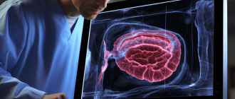
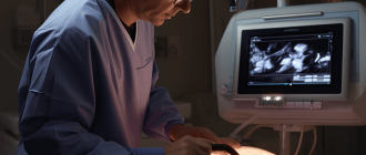
Modern imaging techniques play a vital role in the detection of Median Arcuate Ligament Syndrome. These techniques utilize advanced technologies to capture detailed images of the celiac artery and surrounding structures. The images obtained can provide valuable information to physicians, helping them make an accurate diagnosis.
However, the accuracy of modern imaging in the detection of Median Arcuate Ligament Syndrome is still a subject of debate. While these techniques have greatly improved in recent years, there are still challenges in accurately identifying and evaluating the compression of the celiac artery. False positives and false negatives can occur, leading to misdiagnosis or delayed treatment.
It is important for healthcare professionals to be aware of the limitations of modern imaging in the detection of Median Arcuate Ligament Syndrome. A combination of imaging techniques, clinical evaluation, and patient history should be considered to ensure a reliable and accurate diagnosis. Ongoing research and advancements in imaging technology will continue to improve the accuracy of MALS detection in the future.