CT Scans and MALS: A Deep Dive into Imaging Techniques.
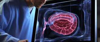
Imaging techniques have taken a deep dive into the world of medical diagnostics, allowing healthcare professionals to visualize the human body in ways that were once unimaginable. CT scans, short for computed tomography scans, have emerged as one of the most powerful tools in this realm. By combining X-ray technology and computer processing, CT scans provide detailed images of internal body structures.
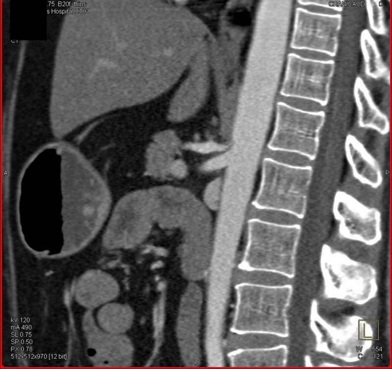
One area where CT scans have proven particularly useful is in the diagnosis and treatment of median arcuate ligament syndrome. MALS is a rare condition that involves the compression of the celiac artery by the median arcuate ligament, resulting in debilitating symptoms. CT scans allow physicians to visualize the anatomy of the abdomen and identify any abnormalities, like the compression of the celiac artery, facilitating accurate diagnosis and appropriate Mals Syndrome Treatment.
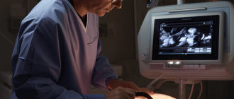
However, the power of CT scans doesn’t stop there. Advanced imaging techniques have been developed to enhance the capabilities of CT scans even further. By leveraging sophisticated algorithms and image processing software, researchers have been able to analyze CT scan data in unprecedented detail. These techniques include 3D reconstruction, which allows physicians to view organs and structures from multiple angles, and virtual colonoscopy, a non-invasive alternative to traditional colonoscopy.
As technology continues to advance, so too will the capabilities of CT scans and other imaging techniques. From identifying tumors at the earliest stages to visualizing blood flow in real-time, the potential of these tools is limitless. By continually exploring and refining these imaging techniques, medical professionals can provide more accurate diagnoses and improve patient outcomes.