
Comparing Diagnostic Tools: CT vs. MRI vs. Ultrasound for MALS.
When it comes to diagnosing a condition called Median Arcuate Ligament Syndrome, various diagnostic tools can be used. Three commonly used tools for diagnosing MALS are computed tomography (CT), magnetic resonance imaging (MRI), and ultrasound. Each of these tools has its strengths and weaknesses, making them suitable for different scenarios and patient populations.
CT scans use X-rays to create detailed images of the body’s internal structures. They are particularly useful for identifying anatomical abnormalities and obtaining precise measurements. CT scans can provide accurate information about the position and compression of the median arcuate ligament, which is the key factor in MALS. This makes CT scans a valuable diagnostic tool for Median Arcuate Ligament Syndrome patients, but after diagnosis, understanding the Mals Syndrome Treatment becomes crucial.
MRI, on the other hand, uses powerful magnets and radio waves to create detailed images of the body’s soft tissues. MRI scans are especially useful for evaluating blood flow to organs and detecting abnormalities in the blood vessels. While CT scans are more focused on anatomical details, MRI provides valuable information about the blood supply to organs affected by Median Arcuate Ligament Syndrome, such as the intestines and kidneys.
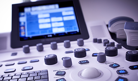
Ultrasound is a non-invasive diagnostic tool that uses sound waves to create images of the body’s internal structures. It is particularly useful for examining blood flow and detecting blockages or abnormalities in the blood vessels. Ultrasound is often used as an initial screening tool for diagnosing MALS due to its affordability, accessibility, and lack of radiation exposure.
Overall, when it comes to diagnosing MALS, a combination of CT, MRI, and ultrasound can provide a comprehensive understanding of the condition. CT scans offer detailed anatomical information, MRI scans provide insights into the blood supply to affected organs, and ultrasound serves as a cost-effective initial screening tool. By utilizing the strengths of each diagnostic tool, healthcare professionals can accurately diagnose and effectively manage Median Arcuate Ligament Syndrome.